Structural investigation of the hedamycin:d(ACCGGT)2 complex by NMR and restrained molecular dynamics.
Owen, E.A., Burley, G.A., Carver, J.A., Wickham, G., Keniry, M.A.(2002) Biochem Biophys Res Commun 290: 1602-1608
- PubMed: 11820806 Search on PubMed
- DOI: https://doi.org/10.1006/bbrc.2002.6369
- Primary Citation Related Structures:
1JHI - PubMed Abstract:
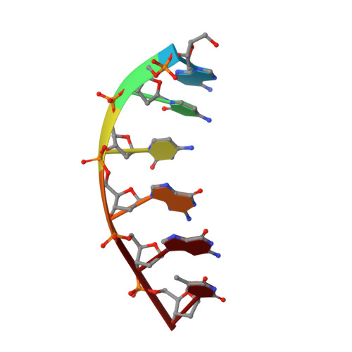
Hedamycin, a member of the pluramycin family of drugs, displays a range of biological responses including antitumor and antimicrobial activity. The mechanism of action is via direct interaction with DNA through intercalation between the bases of the oligonucleotide and alkylation of a guanine residue at 5'-PyG-3' sites. There appears to be some minor structural differences between two earlier studies on the interaction of hedamycin with 5'-PyG-3' sites. In this study, a high-resolution NMR analysis of the hedamycin:d(ACCGGT)2 complex was undertaken in order to investigate the effect of replacing the thymine with a guanine at the preferred 5'-CGT-3' site. The resultant structure was compared with earlier work, with particular emphasis placed on the drug conformation. The structure of the hedamycin:d(ACCGGT)2 complex has many features in common with the two previous NMR structures of hedamycin:DNA complexes but differed in the conformation and orientation of the N,N-dimethylvancosamine saccharide of hedamycin in one of these structures. The preferential binding of hedamycin to 5'-CG-3' over 5'-TG-3' binding sites is explained in terms of the orientation and location of the N,N-dimethylvancosamine saccharide in the minor groove.
- Research School of Chemistry, Australian National University, Canberra, ACT 0200, Australia.
Organizational Affiliation: