1JFD | pdb_00001jfd

STRUCTURE OF INORGANIC PYROPHOSPHATASE


Experimental Data Snapshot

  • Method: X-RAY DIFFRACTION
  • Resolution: 2.20 Å
  • R-Value Free: 
    0.266 (Depositor) 
  • R-Value Work: 
    0.192 (Depositor) 

wwPDB Validation 3D Report Full Report

Validation slider image for 1JFD

This is version 1.3 of the entry. See complete history

Literature

Crystal structure of Escherichia coli inorganic pyrophosphatase complexed with SO4(2-). Ligand-induced molecular asymmetry.

Avaeva, S.Kurilova, S.Nazarova, T.Rodina, E.Vorobyeva, N.Sklyankina, V.Grigorjeva, O.Harutyunyan, E.Oganessyan, V.Wilson, K.Dauter, Z.Huber, R.Mather, T.

(1997) FEBS Lett 410: 502-508

  • DOI: https://doi.org/10.1016/s0014-5793(97)00650-9
  • Primary Citation Related Structures: 
    1JFD

  • PubMed Abstract: 

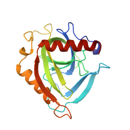
    The three-dimensional structure of inorganic pyrophosphatase from Escherichia coli complexed with sulfate was determined at 2.2 A resolution using Patterson's search technique and refmed to an R-factor of 19.2%. Sulfate may be regarded as a structural analog of phosphate, the product of the enzyme reaction, and as a structural analog of methyl phosphate, the irreversible inhibitor. Sulfate binds to the pyrophosphatase active site cavity as does phosphate and this diminishes molecular symmetry, converting the homohexamer structure form (alpha3)2 into alpha3'alpha3". The asymmetry of the molecule is manifested in displacements of protein functional groups and some parts of the polypeptide chain and reflects the interaction of subunits and their cooperation. The significance of re-arrangements for pyrophosphatase function is discussed.


  • Organizational Affiliation
    • A.N. Belozersky Institute of Physico-Chemical Biology, Moscow State University, Vorobyevy Gory, Russia.

Macromolecule Content 

  • Total Structure Weight: 39.39 kDa 
  • Atom Count: 2,979 
  • Modeled Residue Count: 350 
  • Deposited Residue Count: 350 
  • Unique protein chains: 1

Macromolecules

Find similar proteins by:|  3D Structure
Entity ID: 1
MoleculeChains  Sequence LengthOrganismDetailsImage
INORGANIC PYROPHOSPHATASE
A, B
175Escherichia coliMutation(s): 0 
EC: 3.6.1.1
UniProt
Find proteins for P0A7A9 (Escherichia coli (strain K12))
Explore P0A7A9 
Go to UniProtKB:  P0A7A9
Entity Groups
Sequence Clusters30% Identity50% Identity70% Identity90% Identity95% Identity100% Identity
UniProt GroupP0A7A9
Sequence Annotations
Expand
Reference Sequence

Small Molecules

Experimental Data & Validation

Experimental Data

  • Method: X-RAY DIFFRACTION
  • Resolution: 2.20 Å
  • R-Value Free:  0.266 (Depositor) 
  • R-Value Work:  0.192 (Depositor) 
Space Group: H 3 2
Unit Cell:
Length ( Å )Angle ( ˚ )
a = 110.4α = 90
b = 110.4β = 90
c = 154.8γ = 120
Software Package:
Software NamePurpose
REFMACrefinement
DENZOdata reduction

Structure Validation

View Full Validation Report


Entry History 

Deposition Data

Revision History  (Full details and data files)

  • Version 1.0: 1997-12-03
    Type: Initial release
  • Version 1.1: 2008-03-03
    Changes: Version format compliance
  • Version 1.2: 2011-07-13
    Changes: Derived calculations, Version format compliance
  • Version 1.3: 2024-02-07
    Changes: Data collection, Database references, Derived calculations, Other