1JE8 | pdb_00001je8

Two-Component response regulator NarL/DNA Complex: DNA Bending Found in a High Affinity Site


Experimental Data Snapshot

  • Method: X-RAY DIFFRACTION
  • Resolution: 2.12 Å
  • R-Value Free: 
    0.273 (Depositor) 
  • R-Value Work: 
    0.228 (Depositor), 0.231 (DCC) 

Starting Model: experimental
View more details

wwPDB Validation 3D Report Full Report

Validation slider image for 1JE8

This is version 1.5 of the entry. See complete history

Literature

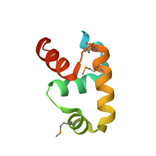
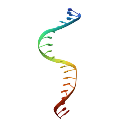
Dimerization allows DNA target site recognition by the NarL response regulator.

Maris, A.E.Sawaya, M.R.Kaczor-Grzeskowiak, M.Jarvis, M.R.Bearson, S.M.Kopka, M.L.Schroder, I.Gunsalus, R.P.Dickerson, R.E.

(2002) Nat Struct Biol 9: 771-778

  • DOI: https://doi.org/10.1038/nsb845
  • Primary Citation Related Structures: 
    1JE8

  • PubMed Abstract: 

    Two-component signal transduction systems are modular phosphorelay regulatory pathways common in prokaryotes. In the co-crystal structure of the Escherichia coli NarL signal output domain bound to DNA, we observe how the NarL family of two-component response regulators can bind DNA. DNA recognition is accompanied by the formation of a new dimerization interface, which could occur only in the full-length protein via a large intramolecular domain rearrangement. The DNA is recognized by the concerted effects of solvation, van der Waals forces and inherent DNA deformability, rather than determined primarily by major groove hydrogen bonding. These subtle forces permit a small DNA-binding domain to perturb the DNA helix, leading to major DNA curvature and a transition from B- to A-form DNA at the binding site, where valine on the recognition helix interacts unexpectedly with the polar major groove floor.


  • Organizational Affiliation
    • Department of Chemistry and Biochemistry, University of California, Los Angeles, P.O. Box 951569, Los Angeles, California 90095-1569, USA.

Macromolecule Content 

  • Total Structure Weight: 64.62 kDa 
  • Atom Count: 4,199 
  • Modeled Residue Count: 345 
  • Deposited Residue Count: 408 
  • Unique protein chains: 1
  • Unique nucleic acid chains: 1

Macromolecules


Find similar proteins by:|  3D Structure
Entity ID: 2
MoleculeChains  Sequence LengthOrganismDetailsImage
Nitrate/Nitrite Response Regulator Protein NARLE [auth A],
F [auth B],
G [auth E],
H [auth F]
82Escherichia coliMutation(s): 3 
UniProt
Find proteins for P0AF28 (Escherichia coli (strain K12))
Explore P0AF28 
Go to UniProtKB:  P0AF28
Entity Groups
Sequence Clusters30% Identity50% Identity70% Identity90% Identity95% Identity100% Identity
UniProt GroupP0AF28
Sequence Annotations
Expand
Reference Sequence
Find similar nucleic acids by:  Sequence
Entity ID: 1
MoleculeChains LengthOrganismImage
5'-D(*CP*GP*TP*AP*CP*CP*CP*AP*TP*TP*AP*AP*TP*GP*GP*GP*TP*AP*CP*G)-3'A [auth C],
B [auth D],
C [auth G],
D [auth H]
20N/A
Sequence Annotations
Expand
Reference Sequence

Small Molecules

Modified Residues  1 Unique
IDChains TypeFormula2D DiagramParent
MSE
Query on MSE
E [auth A],
F [auth B],
G [auth E],
H [auth F]
L-PEPTIDE LINKINGC5 H11 N O2 SeMET

Experimental Data & Validation

Experimental Data

  • Method: X-RAY DIFFRACTION
  • Resolution: 2.12 Å
  • R-Value Free:  0.273 (Depositor) 
  • R-Value Work:  0.228 (Depositor), 0.231 (DCC) 
Space Group: P 1 21 1
Unit Cell:
Length ( Å )Angle ( ˚ )
a = 76.257α = 90
b = 52.741β = 90
c = 83.863γ = 90
Software Package:
Software NamePurpose
DENZOdata reduction
TRUNCATEdata reduction
GLRFphasing
CNSrefinement
CCP4data scaling

Structure Validation

View Full Validation Report


Entry History 

Revision History  (Full details and data files)

  • Version 1.0: 2002-09-27
    Type: Initial release
  • Version 1.1: 2008-04-27
    Changes: Version format compliance
  • Version 1.2: 2011-07-13
    Changes: Version format compliance
  • Version 1.3: 2023-08-16
    Changes: Data collection, Database references, Derived calculations, Refinement description
  • Version 1.4: 2023-11-15
    Changes: Data collection
  • Version 1.5: 2024-10-09
    Changes: Structure summary