Structural comparison of monomeric variants of the chemokine MIP-1beta having differing ability to bind the receptor CCR5.
Kim, S., Jao, S., Laurence, J.S., LiWang, P.J.(2001) Biochemistry 40: 10782-10791
- PubMed: 11535053 Search on PubMed
- DOI: https://doi.org/10.1021/bi011065x
- Primary Citation Related Structures:
1JE4 - PubMed Abstract:
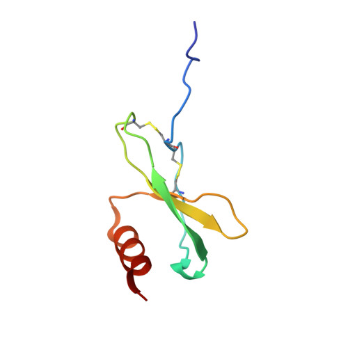
MIP-1beta, a member of the chemokine family of proteins, tightly binds the receptor CCR5 as part of its natural function in the immune response, and in doing so also blocks the ability of many strains of HIV to enter the cell. The single most important MIP-1beta residue known to contribute to its interaction with the receptor is Phe13, which when mutated reduces the ability of MIP-1beta to bind to CCR5 by more than 1000-fold. To obtain a structural understanding of the dramatic effect of the absence of Phe13 in MIP-1beta, we used multidimensional heteronuclear NMR to determine the three-dimensional structure of the MIP-1beta F13A variant. We had previously shown that, unlike the wild-type protein which has been shown to be a tight dimer, the F13A mutant is monomeric even at high concentrations [Laurence, J. S., Blanpain, C., Burgner, J. W., Parmentier, M., and LiWang, P. J. (2000) Biochemistry 39, 3401-3409], leading to significant changes in the NMR spectra of F13A and the wild-type protein. We have obtained a total of 940 structural restraints for MIP-1beta F13A, and have calculated a family of structures having a backbone rmsd from the average of 0.55 A (residues 12-67). A structural comparison of the F13A mutant with a fully active monomeric variant, P8A, shows that despite some differences in the (1)H-(15)N HSQC spectra the two are nearly identical in NOE distance restraints and in backbone conformation. A comparison of F13A with the wild-type protein shows largely the same fold, although differences exist in the N-terminal and loop regions for which the loss of the dimer in F13A can mainly account. A dynamics comparison confirms greater flexibility in F13A than in the wild-type protein in regions of dimer contact in the wild-type protein. In an analysis to determine if the large functional effect resulting from the loss of Phe13 is due to the local side chain change or due to more global structural changes, we conclude that local effects predominate. This suggests that a strategy for designing tight binding anti-CCR5 therapeutics should include a Phe-like component.
- Department of Biochemistry and Biophysics, Texas A&M University, TAMU 2128, College Station, Texas 77843-2128, USA.
Organizational Affiliation: