1JDC | pdb_00001jdc

MUTANT (E219Q) MALTOTETRAOSE-FORMING EXO-AMYLASE COCRYSTALLIZED WITH MALTOTETRAOSE (CRYSTAL TYPE 1)


Experimental Data Snapshot

  • Method: X-RAY DIFFRACTION
  • Resolution: 1.90 Å
  • R-Value Work: 
    0.174 (Depositor), 0.164 (DCC) 

wwPDB Validation 3D Report Full Report

Validation slider image for 1JDC

This is version 2.2 of the entry. See complete history

Literature

Crystal structures of a mutant maltotetraose-forming exo-amylase cocrystallized with maltopentaose.

Yoshioka, Y.Hasegawa, K.Matsuura, Y.Katsube, Y.Kubota, M.

(1997) J Mol Biology 271: 619-628

  • DOI: https://doi.org/10.1006/jmbi.1997.1222
  • Primary Citation Related Structures: 
    1JDA, 1JDC, 1JDD

  • PubMed Abstract: 

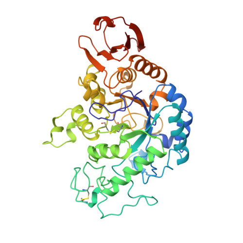
    The three-dimensional structures of the catalytic residue Glu219-->Gln mutant of Pseudomonas stutzeri maltotetraose-forming exo-alpha-amylase, and its complex with carbohydrate obtained by cocrystallization with maltopentaose were determined. Two crystal forms were obtained for the complexed enzyme, and a bound maltotetraose was found in each. The structures were analyzed at 2.2 A and 1.9 A resolution, respectively for the uncomplexed and complexed mutant. These structures were compared with the wild-type enzyme structure. In the complexed crystals, the maltotetraose was firmly bound, extensively interacting with the amino acid environments in the active cleft. The non-reducing end glucose unit was hydrogen bonded to the side-chain of Asp160 and the main-chain nitrogen of Gly158, which seem to be predominantly required for the recognition of the non-reducing end of the substrate that determines the exo-wise degradation of this enzyme. The reducing end glucose unit of bound maltotetraose showed clear deformation, adopting a half-chair conformation with extensive hydrogen bonds to surrounding polypeptides. The C1-atom of this deformed glucose unit lies very close to Asp193OD1 with a distance of 2.6 A. The catalytic residue Asp294 is firmly hydrogen-bonded to the O2 and O3-hydroxyl groups of the deformed reducing end glucose unit. Upon binding of the carbohydrate, small but significant induced fits were observed in the regions of Asp294, Phe156, Ile157, and Asp160. Possible roles of the three catalytic residues are also discussed.


  • Organizational Affiliation
    • Institute for Protein Research, Osaka University, Osaka, Suita, 565, Japan.

Macromolecule Content 

  • Total Structure Weight: 48.16 kDa 
  • Atom Count: 3,556 
  • Modeled Residue Count: 418 
  • Deposited Residue Count: 429 
  • Unique protein chains: 1

Macromolecules

Find similar proteins by:|  3D Structure
Entity ID: 1
MoleculeChains  Sequence LengthOrganismDetailsImage
1,4-ALPHA MALTOTETRAHYDROLASE429Stutzerimonas stutzeriMutation(s): 1 
EC: 3.2.1.60
UniProt
Find proteins for P13507 (Stutzerimonas stutzeri)
Explore P13507 
Go to UniProtKB:  P13507
Entity Groups
Sequence Clusters30% Identity50% Identity70% Identity90% Identity95% Identity100% Identity
UniProt GroupP13507
Sequence Annotations
Expand
Reference Sequence

Oligosaccharides

Help  
Entity ID: 2
MoleculeChains Length2D Diagram GlycosylationD Interactions
alpha-D-glucopyranose-(1-4)-alpha-D-glucopyranose-(1-4)-alpha-D-glucopyranose-(1-4)-alpha-D-glucopyranose
B
4N/A
Glycosylation Resources
GlyTouCan: G87171PZ
GlyCosmos: G87171PZ
GlyGen: G87171PZ

Biologically Interesting Molecules (External Reference) 

1 Unique

Experimental Data & Validation

Experimental Data

  • Method: X-RAY DIFFRACTION
  • Resolution: 1.90 Å
  • R-Value Work:  0.174 (Depositor), 0.164 (DCC) 
Space Group: P 21 21 21
Unit Cell:
Length ( Å )Angle ( ˚ )
a = 65.1α = 90
b = 138.4β = 90
c = 50.7γ = 90
Software Package:
Software NamePurpose
X-PLORmodel building
PROFFTrefinement
X-PLORrefinement
X-PLORphasing

Structure Validation

View Full Validation Report


Entry History 

Deposition Data

Revision History  (Full details and data files)

  • Version 1.0: 1997-10-15
    Type: Initial release
  • Version 1.1: 2008-03-24
    Changes: Version format compliance
  • Version 1.2: 2011-07-13
    Changes: Version format compliance
  • Version 2.0: 2020-07-29
    Type: Remediation
    Reason: Carbohydrate remediation
    Changes: Atomic model, Data collection, Database references, Derived calculations, Other, Structure summary
  • Version 2.1: 2021-11-03
    Changes: Database references, Structure summary
  • Version 2.2: 2024-10-30
    Changes: Data collection, Structure summary