The dimerization domain of HNF-1alpha: structure and plasticity of an intertwined four-helix bundle with application to diabetes mellitus.
Narayana, N., Hua, Q., Weiss, M.A.(2001) J Mol Biol 310: 635-658
- PubMed: 11439029 Search on PubMed
- DOI: https://doi.org/10.1006/jmbi.2001.4780
- Primary Citation Related Structures:
1JB6 - PubMed Abstract:
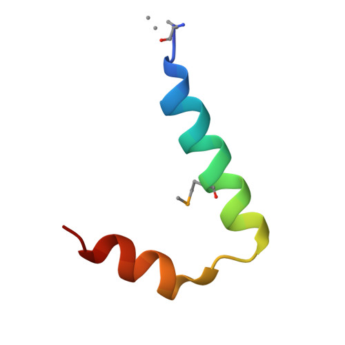
Maturity-onset diabetes mellitus of the young (MODY) is a human genetic syndrome most commonly due to mutations in hepatocyte nuclear factor-1alpha (HNF-1alpha). Here, we describe the crystal structure of the HNF-1alpha dimerization domain at 1.7 A resolution and assess its structural plasticity. The crystal's low solvent content (23%, v/v) leads to tight packing of peptides in the lattice. Two independent dimers, similar in structure, are formed in the unit cell by a 2-fold crystallographic symmetry axis. The dimers define a novel intertwined four-helix bundle (4HB). Each protomer contains two alpha-helices separated by a sharp non-canonical turn. Dimer-related alpha-helices form anti-parallel coiled-coils, including an N-terminal "mini-zipper" complementary in structure, symmetry and surface characteristics to transcriptional coactivator dimerization cofactor of HNF-1 (DCoH). A confluence of ten leucine side-chains (five per protomer) forms a hydrophobic core. Isotope-assisted NMR studies demonstrate that a similar intertwined dimer exists in solution. Comparison of structures obtained in multiple independent crystal forms indicates that the mini-zipper is a stable structural element, whereas the C-terminal alpha-helix can adopt a broad range of orientations. Segmental alignment of the mini-zipper (mean pairwise root-mean-square difference (rmsd) in C(alpha) coordinates of 0.29 A) is associated with a 2.1 A mean C(alpha) rmsd displacement of the C-terminal coiled-coil. The greatest C-terminal structural variation (4.1 A C(alpha) rmsd displacement) is observed in the DCoH-bound peptide. Diabetes-associated mutations perturb distinct structural features of the HNF-1alpha domain. One mutation (L12H) destabilizes the domain but preserves structural specificity. Adjoining H12 side-chains in a native-like dimer are predicted to alter the functional surface of the mini-zipper involved in DCoH recognition. The other mutation (G20R), by contrast, leads to a dimeric molten globule, as indicated by its 1H-NMR features and fluorescent binding of 1-anilino-8-naphthalene sulfonate. We propose that a glycine-specific turn configuration enables specific interactions between the mini-zipper and the C-terminal coiled-coil.
- Department of Biochemistry, Case Western Reserve University, 10900 Euclid Avenue, Cleveland, OH 44106-4935, USA.
Organizational Affiliation: