1JB3

The Laminin-Binding Domain of Agrin is structurally related to N-TIMP-1


Experimental Data Snapshot

  • Method: X-RAY DIFFRACTION
  • Resolution: 1.60 Å
  • R-Value Free: 0.243 
  • R-Value Work: 0.198 
  • R-Value Observed: 0.198 

wwPDB Validation   3D Report Full Report


This is version 1.3 of the entry. See complete history


Literature

The laminin-binding domain of agrin is structurally related to N-TIMP-1.

Stetefeld, J.Jenny, M.Schulthess, T.Landwehr, R.Schumacher, B.Frank, S.Ruegg, M.A.Engel, J.Kammerer, R.A.

(2001) Nat Struct Biol 8: 705-709

  • DOI: https://doi.org/10.1038/90422
  • Primary Citation of Related Structures:  
    1JB3, 1JC7

  • PubMed Abstract: 

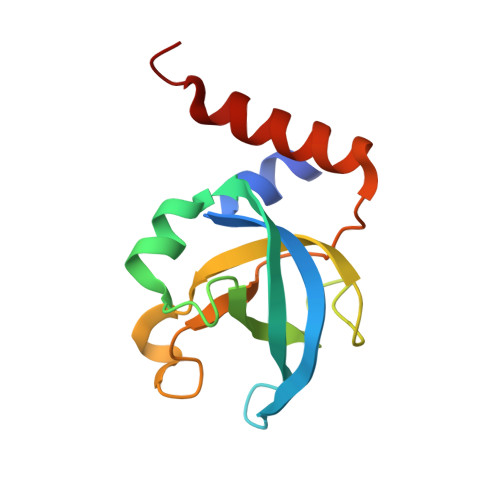
    Agrin is the key organizer of postsynaptic differentiation at the neuromuscular junction. This organization activity requires the binding of agrin to the synaptic basal lamina. Binding is conferred by the N-terminal agrin (NtA) domain, which mediates a high-affinity interaction with the coiled coil domain of laminins. Here, we report the crystal structure of chicken NtA at 1.6 A resolution. The structure reveals that NtA harbors an oligosaccharide/oligonucleotide-binding fold with several possible sites for the interaction with different ligands. A high structural similarity of NtA with the protease inhibition domain in tissue inhibitor of metalloproteinases-1 (TIMP-1) supports the idea of additional functions of agrin besides synaptogenic activity.


  • Organizational Affiliation

    Department of Biophysical Chemistry, Biozentrum, University of Basel, Klingelbergstrasse 70, CH-4056 Basel, Switzerland.


Macromolecules
Find similar proteins by:  (by identity cutoff)  |  3D Structure
Entity ID: 1
MoleculeChains Sequence LengthOrganismDetailsImage
Agrin131Gallus gallusMutation(s): 0 
UniProt
Find proteins for P31696 (Gallus gallus)
Explore P31696 
Go to UniProtKB:  P31696
Entity Groups  
Sequence Clusters30% Identity50% Identity70% Identity90% Identity95% Identity100% Identity
UniProt GroupP31696
Sequence Annotations
Expand
  • Reference Sequence
Experimental Data & Validation

Experimental Data

  • Method: X-RAY DIFFRACTION
  • Resolution: 1.60 Å
  • R-Value Free: 0.243 
  • R-Value Work: 0.198 
  • R-Value Observed: 0.198 
  • Space Group: C 1 2 1
Unit Cell:
Length ( Å )Angle ( ˚ )
a = 76.13α = 90
b = 49.46β = 116.4
c = 53.28γ = 90
Software Package:
Software NamePurpose
SHARPphasing
CNSrefinement
MOSFLMdata reduction
CCP4data scaling

Structure Validation

View Full Validation Report


Entry History 

Deposition Data

Revision History  (Full details and data files)

  • Version 1.0: 2001-08-08
    Type: Initial release
  • Version 1.1: 2008-04-27
    Changes: Version format compliance
  • Version 1.2: 2011-07-13
    Changes: Version format compliance
  • Version 1.3: 2024-02-07
    Changes: Data collection, Database references