1J7X | pdb_00001j7x

CRYSTAL STRUCTURE OF A FUNCTIONAL UNIT OF INTERPHOTORECEPTOR RETINOID-BINDING PROTEIN (IRBP)


Experimental Data Snapshot

  • Method: X-RAY DIFFRACTION
  • Resolution: 1.80 Å
  • R-Value Free: 
    0.220 (Depositor) 
  • R-Value Work: 
    0.205 (Depositor) 
  • R-Value Observed: 
    0.207 (Depositor) 

wwPDB Validation 3D Report Full Report

Validation slider image for 1J7X

This is version 1.3 of the entry. See complete history

Literature

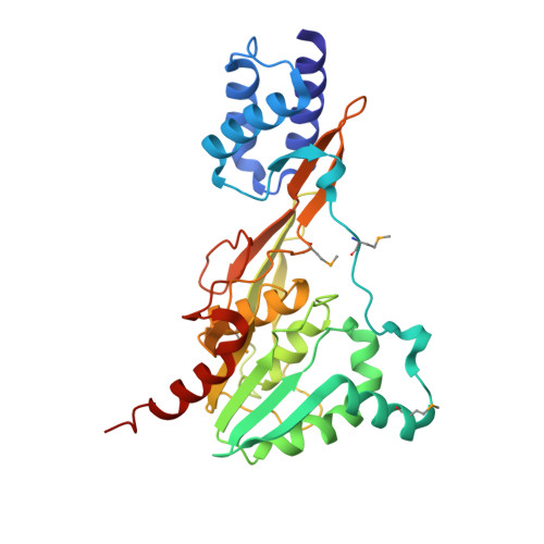
Crystal structure of the functional unit of interphotoreceptor retinoid binding protein.

Loew, A.Gonzalez-Fernandez, F.

(2002) Structure 10: 43-49

  • DOI: https://doi.org/10.1016/s0969-2126(01)00698-0
  • Primary Citation Related Structures: 
    1J7X

  • PubMed Abstract: 

    Interphotoreceptor retinoid binding protein (IRBP), the major soluble component of the interphotoreceptor matrix, is critical to the function, integrity, and development of the vertebrate retina. Although its role is poorly understood, IRBP has been thought to protect 11-cis retinal and all-trans retinol while facilitating their exchange between the photoreceptors and retinal-pigmented epithelium. We determined the X-ray structure of one of the functional units, or modules, of Xenopus laevis IRBP to 1.8 A resolution by multiwavelength anomalous dispersion. The monomeric protein consists of two domains separated by a hydrophobic ligand binding site. A structural homology to the recently solved photosystem II D1 C-terminal-processing protease and the enoyl-CoA isomerase/hydratase family suggests the utility of a common fold used in diverse settings, ranging from proteolysis to fatty acid isomerization to retinoid transport.


  • Organizational Affiliation
    • Department of Biochemistry, University of Texas, Southwestern Medical Center at Dallas, 5323 Harry Hines Boulevard, Dallas, TX 75390, USA. andreas.loew@abbott.com

Macromolecule Content 

  • Total Structure Weight: 33.51 kDa 
  • Atom Count: 2,485 
  • Modeled Residue Count: 302 
  • Deposited Residue Count: 302 
  • Unique protein chains: 1

Macromolecules

Find similar proteins by:|  3D Structure
Entity ID: 1
MoleculeChains  Sequence LengthOrganismDetailsImage
INTERPHOTORECEPTOR RETINOID-BINDING PROTEIN302Xenopus laevisMutation(s): 0 
UniProt
Find proteins for Q7SZI7 (Xenopus laevis)
Explore Q7SZI7 
Go to UniProtKB:  Q7SZI7
Entity Groups
Sequence Clusters30% Identity50% Identity70% Identity90% Identity95% Identity100% Identity
UniProt GroupQ7SZI7
Sequence Annotations
Expand
Reference Sequence

Small Molecules

Modified Residues  1 Unique
IDChains TypeFormula2D DiagramParent
MSE
Query on MSE
A
L-PEPTIDE LINKINGC5 H11 N O2 SeMET

Experimental Data & Validation

Experimental Data

  • Method: X-RAY DIFFRACTION
  • Resolution: 1.80 Å
  • R-Value Free:  0.220 (Depositor) 
  • R-Value Work:  0.205 (Depositor) 
  • R-Value Observed: 0.207 (Depositor) 
Space Group: P 21 21 21
Unit Cell:
Length ( Å )Angle ( ˚ )
a = 63.144α = 90
b = 65.756β = 90
c = 68.174γ = 90
Software Package:
Software NamePurpose
SOLVEphasing
REFMACrefinement
DENZOdata reduction
SCALEPACKdata scaling

Structure Validation

View Full Validation Report


Entry History 

Deposition Data

Revision History  (Full details and data files)

  • Version 1.0: 2002-02-20
    Type: Initial release
  • Version 1.1: 2008-04-27
    Changes: Version format compliance
  • Version 1.2: 2011-07-13
    Changes: Version format compliance
  • Version 1.3: 2024-11-13
    Changes: Data collection, Database references, Derived calculations, Structure summary