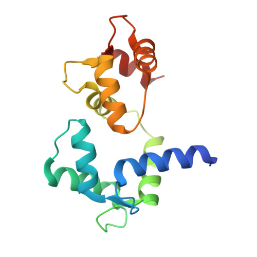
Target-induced conformational adaptation of calmodulin revealed by the crystal structure of a complex with nematode Ca(2+)/calmodulin-dependent kinase kinase peptide
Kurokawa, H., Osawa, M., Kurihara, H., Katayama, N., Tokumitsu, H., Swindells, M.B., Kainosho, M., Ikura, M.(2001) J Mol Biology 312: 59-68
- PubMed: 11545585
- DOI: https://doi.org/10.1006/jmbi.2001.4822
- Primary Citation of Related Structures:
1IQ5 - PubMed Abstract:
Calmodulin (CaM) is a ubiquitous calcium (Ca(2+)) sensor which binds and regulates protein serine/threonine kinases along with many other proteins in a Ca(2+)-dependent manner. For this multi-functionality, conformational plasticity is essential; however, the nature and magnitude of CaM's plasticity still remains largely undetermined. Here, we present the 1.8 A resolution crystal structure of Ca(2+)/CaM, complexed with the 27-residue synthetic peptide corresponding to the CaM-binding domain of the nematode Caenorhabditis elegans Ca(2+)/CaM-dependent kinase kinase (CaMKK). The peptide bound in this crystal structure is a homologue of the previously NMR-derived complex with rat CaMKK, but benefits from improved structural resolution. Careful comparison of the present structure to previous crystal structures of CaM complexed with unrelated peptides derived from myosin light chain kinase and CaM kinase II, allow a quantitative analysis of the differences in the relative orientation of the N and C-terminal domains of CaM, defined as a screw axis rotation angle ranging from 156 degrees to 196 degrees. The principal differences in CaM interaction with various peptides are associated with the N-terminal domain of CaM. Unlike the C-terminal domain, which remains unchanged internally, the N-terminal domain of CaM displays significant differences in the EF-hand helix orientation between this and other CaM structures. Three hydrogen bonds between CaM and the peptide (E87-R336, E87-T339 and K75-T339) along with two salt bridges (E11-R349 and E114-K334) are the most probable determinants for the binding direction of the CaMKK peptide to CaM.
- Division of Molecular and Structural Biology, Ontario Cancer Institute and Department of Medical Biophysics, University of Toronto, Ontario M5G2M9, Canada.
Organizational Affiliation:


