1IOK | pdb_00001iok

CRYSTAL STRUCTURE OF CHAPERONIN-60 FROM PARACOCCUS DENITRIFICANS


Experimental Data Snapshot

  • Method: X-RAY DIFFRACTION
  • Resolution: 3.20 Å
  • R-Value Free: 
    0.235 (Depositor) 
  • R-Value Work: 
    0.204 (Depositor) 
  • R-Value Observed: 
    0.203 (Depositor) 

wwPDB Validation 3D Report Full Report

Validation slider image for 1IOK

This is version 1.3 of the entry. See complete history

Literature

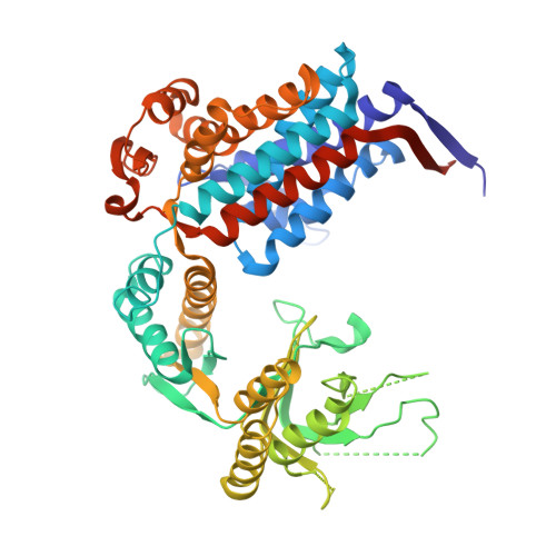
Crystal structure of chaperonin-60 from Paracoccus denitrificans.

Fukami, T.A.Yohda, M.Taguchi, H.Yoshida, M.Miki, K.

(2001) J Mol Biology 312: 501-509

  • DOI: https://doi.org/10.1006/jmbi.2001.4961
  • Primary Citation Related Structures: 
    1IOK

  • PubMed Abstract: 

    The crystal structure of chaperonin-60 from Paracoccus denitrificans (P.cpn60) has been determined at 3.2 A resolution by the molecular replacement method. Two heptameric rings of identical subunits of P.cpn60 in adjacent asymmetric units are stacked in a back-to-back manner and form a cylinder, as found in GroEL, cpn60 from Escherichia coli. With respect to the unliganded GroEL structure, each subunit of P.cpn60 tilts 2 degrees outwards and the apical domain twists 4 degrees counter-clockwise in the top view in a hinge-like manner, rendering the central hole 5 A wider. Despite the subunit tilts, both rings in P.cpn60 contact at two sites of the equatorial domain in the same way as in GroEL. Interactions between residues 434 and 434, and 463 and 463 observed in GroEL were not found in P.cpn60, and the interaction between 452 and 461 was weaker in P.cpn60 than in GroEL. The unique hydrogen bond between 468 and 471 was observed at the right site in P.cpn60, which could account for why the subunits tilt outwards. The contact surface area was reduced at the left site, which is similar to the observed changes in the GroEL structures induced by ATP binding. In general, inter-ring interactions in P.cpn60 were weakened, which is consistent with findings that P.cpn60 is observed in single-ring forms as well as in double-ring forms.


  • Organizational Affiliation
    • Department of Chemistry Graduate School of Science, Kyoto University, Kyoto, Sakyo-ku, 606-8502, Japan.

Macromolecule Content 

  • Total Structure Weight: 404.52 kDa 
  • Atom Count: 25,095 
  • Modeled Residue Count: 3,374 
  • Deposited Residue Count: 3,815 
  • Unique protein chains: 1

Macromolecules

Find similar proteins by:|  3D Structure
Entity ID: 1
MoleculeChains  Sequence LengthOrganismDetailsImage
CHAPERONIN 60
A, B, C, D, E
A, B, C, D, E, F, G
545Paracoccus denitrificansMutation(s): 0 
EC: 5.6.1.7
UniProt
Find proteins for Q9Z462 (Paracoccus denitrificans)
Explore Q9Z462 
Go to UniProtKB:  Q9Z462
Entity Groups
Sequence Clusters30% Identity50% Identity70% Identity90% Identity95% Identity100% Identity
UniProt GroupQ9Z462
Sequence Annotations
Expand
Reference Sequence

Experimental Data & Validation

Experimental Data

  • Method: X-RAY DIFFRACTION
  • Resolution: 3.20 Å
  • R-Value Free:  0.235 (Depositor) 
  • R-Value Work:  0.204 (Depositor) 
  • R-Value Observed: 0.203 (Depositor) 
Space Group: P 42 21 2
Unit Cell:
Length ( Å )Angle ( ˚ )
a = 286.355α = 90
b = 286.355β = 90
c = 153.463γ = 90
Software Package:
Software NamePurpose
X-PLORmodel building
X-PLORrefinement
DENZOdata reduction
SCALEPACKdata scaling
X-PLORphasing

Structure Validation

View Full Validation Report


Entry History 

Deposition Data

Revision History  (Full details and data files)

  • Version 1.0: 2001-10-03
    Type: Initial release
  • Version 1.1: 2008-04-27
    Changes: Version format compliance
  • Version 1.2: 2011-07-13
    Changes: Version format compliance
  • Version 1.3: 2023-12-27
    Changes: Data collection, Database references