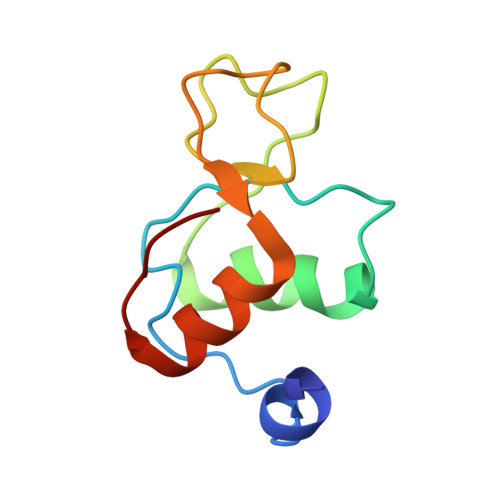
Solution structure and backbone dynamics of the human DNA ligase IIIalpha BRCT domain
Krishnan, V.V., Thornton, K.H., Thelen, M.P., Cosman, M.(2001) Biochemistry 40: 13158-13166
- PubMed: 11683624
- DOI: https://doi.org/10.1021/bi010979g
- Primary Citation of Related Structures:
1IMO, 1IN1 - PubMed Abstract:
BRCT (BRCA1 carboxyl terminus) domains are found in a number of DNA repair enzymes and cell cycle regulators and are believed to mediate important protein-protein interactions. The DNA ligase IIIalpha BRCT domain partners with the distal BRCT domain of the DNA repair protein XRCC1 (X1BRCTb) in the DNA base excision repair (BER) pathway. To elucidate the mechanisms by which these two domains can interact, we have determined the solution structure of human ligase IIIalpha BRCT (L3[86], residues 837-922). The structure of L3[86] consists of a beta2beta1beta3beta4 parallel sheet with a two-alpha-helix bundle packed against one face of the sheet. This fold is conserved in several proteins having a wide range of activities, including X1BRCTb [Zhang, X. D., et al. (1998) EMBO J. 17, 6404-6411]. L3[86] exists as a dimer in solution, but an insufficient number of NOE restraints precluded the determination of the homodimer structure. However, 13C isotope-filtered and hydrogen-deuterium exchange experiments indicate that the N-terminus, alpha1, the alpha1-beta2 loop, and the three residues following alpha2 are involved in forming the dimer interface, as similarly observed in the structure of X1BRCTb. NOE and dynamic data indicate that several residues (837-844) in the N-terminal region appear to interconvert between helix and random coil conformations. Further studies of other BRCT domains and of their complexes are needed to address how these proteins interact with one another, and to shed light on how mutations can lead to disruption of function and ultimately disease.
- Biology and Biotechnology Research Program, Lawrence Livermore National Laboratory, Livermore, California 94551, USA.
Organizational Affiliation:


