1ILQ

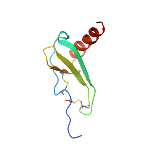
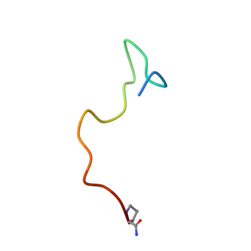
CXCR-1 N-TERMINAL PEPTIDE BOUND TO INTERLEUKIN-8 (MINIMIZED MEAN)


Experimental Data Snapshot

  • Method: SOLUTION NMR
  • Conformers Calculated: 40 
  • Conformers Submitted: 
  • Selection Criteria: LEAST RESTRAINT VIOLATION ENERGY 

wwPDB Validation   3D Report Full Report


This is version 2.1 of the entry. See complete history


Literature

Structure of a CXC chemokine-receptor fragment in complex with interleukin-8.

Skelton, N.J.Quan, C.Reilly, D.Lowman, H.

(1999) Structure 7: 157-168

  • DOI: https://doi.org/10.1016/S0969-2126(99)80022-7

  • PubMed Abstract: 

    Interactions between CXC chemokines (e.g. interleukin-8, IL-8) and their receptors (e.g. CXCR-1) have a key role in host defense and disease by attracting and upregulating neutrophils to sites of inflammation. The transmembrane nature of the receptor impedes structure-based understanding of ligand interactions. Linear peptides based on the N-terminal, extracellular portion of the receptor CXCR-1 do bind to IL-8, however, and inhibit the binding of IL-8 to the full-length receptor. The NMR solution structure of the complex formed between IL-8 and one such receptor-based peptide indicates that a cleft between a loop and a beta hairpin constitute part of the receptor interaction surface on IL-8. Nine residues from the C terminus of the receptor peptide (corresponding to Pro21-Pro29 of CXCR-1) occupy the cleft in an extended fashion. Intermolecular contacts are mostly hydrophobic and sidechain mediated. The results offer the first details at an atomic level of the interaction between a chemokine and its receptor. Consideration of other biochemical data allow extrapolation to a model for the interaction of IL-8 with the full-length receptor. In this model, the heparin-binding residues of IL-8 are exposed, thereby allowing presentation of the chemokine from endothelial cell-surface glycosaminoglycans. This first glimpse of how IL-8 binds to its receptor provides a foundation for the structure-based design of chemokine antagonists.


  • Organizational Affiliation

    Department of Protein Engineering Genentech, Inc. South San Francisco, CA 94080, USA. skelly@gene.com


Macromolecules
Find similar proteins by:  (by identity cutoff)  |  3D Structure
Entity ID: 1
MoleculeChains Sequence LengthOrganismDetailsImage
INTERLEUKIN-8 PRECURSOR
A, B
72Homo sapiensMutation(s): 0 
UniProt & NIH Common Fund Data Resources
Find proteins for P10145 (Homo sapiens)
Explore P10145 
Go to UniProtKB:  P10145
PHAROS:  P10145
GTEx:  ENSG00000169429 
Entity Groups  
Sequence Clusters30% Identity50% Identity70% Identity90% Identity95% Identity100% Identity
UniProt GroupP10145
Sequence Annotations
Expand
  • Reference Sequence

Find similar proteins by:  Sequence   |   3D Structure  

Entity ID: 2
MoleculeChains Sequence LengthOrganismDetailsImage
INTERLEUKIN-8 RECEPTOR A19Homo sapiensMutation(s): 0 
UniProt & NIH Common Fund Data Resources
Find proteins for P25024 (Homo sapiens)
Explore P25024 
Go to UniProtKB:  P25024
PHAROS:  P25024
GTEx:  ENSG00000163464 
Entity Groups  
Sequence Clusters30% Identity50% Identity70% Identity90% Identity95% Identity100% Identity
UniProt GroupP25024
Sequence Annotations
Expand
  • Reference Sequence
Experimental Data & Validation

Experimental Data

  • Method: SOLUTION NMR
  • Conformers Calculated: 40 
  • Conformers Submitted: 
  • Selection Criteria: LEAST RESTRAINT VIOLATION ENERGY 

Structure Validation

View Full Validation Report


Entry History 

Deposition Data

Revision History  (Full details and data files)

  • Version 1.0: 1998-12-23
    Type: Initial release
  • Version 1.1: 2008-03-24
    Changes: Version format compliance
  • Version 1.2: 2011-07-13
    Changes: Version format compliance
  • Version 1.3: 2016-05-25
    Changes: Source and taxonomy
  • Version 1.4: 2022-02-23
    Changes: Data collection, Database references, Derived calculations, Other
  • Version 2.0: 2023-11-15
    Changes: Atomic model, Data collection, Derived calculations
  • Version 2.1: 2024-10-23
    Changes: Structure summary