1II0 | pdb_00001ii0

CRYSTAL STRUCTURE OF THE ESCHERICHIA COLI ARSENITE-TRANSLOCATING ATPASE


Experimental Data Snapshot

  • Method: X-RAY DIFFRACTION
  • Resolution: 2.40 Å
  • R-Value Free: 
    0.252 (Depositor), 0.246 (DCC) 
  • R-Value Work: 
    0.210 (Depositor), 0.204 (DCC) 
  • R-Value Observed: 
    0.210 (Depositor) 

wwPDB Validation 3D Report Full Report

Validation slider image for 1II0

Ligand Structure Quality Assessment 


This is version 1.4 of the entry. See complete history

Literature

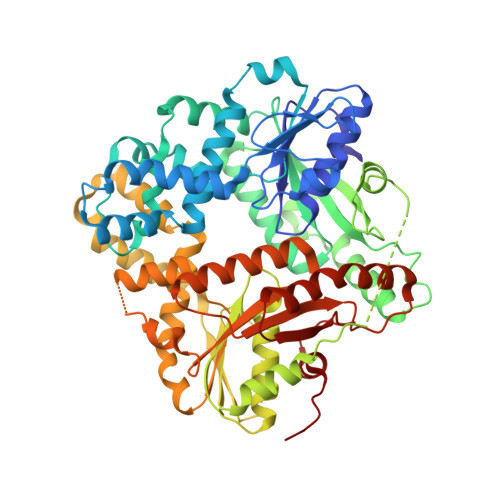
Conformational changes in four regions of the Escherichia coli ArsA ATPase link ATP hydrolysis to ion translocation.

Zhou, T.Radaev, S.Rosen, B.P.Gatti, D.L.

(2001) J Biological Chem 276: 30414-30422

  • DOI: https://doi.org/10.1074/jbc.M103671200
  • Primary Citation Related Structures: 
    1IHU, 1II0, 1II9

  • PubMed Abstract: 

    Structures of ArsA with ATP, AMP-PNP, or ADP.AlF(3) bound at the A2 nucleotide binding site were determined. Binding of different nucleotides modifies the coordination sphere of Mg(2+). In particular, the changes elicited by ADP.AlF(3) provide insights into the mechanism of ATP hydrolysis. In-line attack by water onto the gamma-phosphate of ATP would be followed first by formation of a trigonal intermediate and then by breaking of the scissile bond between the beta- and gamma-phosphates. Motions of amino acid side chains at the A2 nucleotide binding site during ATP binding and hydrolysis propagate at a distance, producing conformational changes in four different regions of the protein corresponding to helices H4-H5, helices H9-H10, helices H13-H15, and to the S1-H2-S2 region. These elements are extensions of, respectively, the Switch I and Switch II regions, the A-loop (a small loop near the nucleotide adenine moiety), and the P-loop. Based on the observed conformational changes, it is proposed that ArsA functions as a reciprocating engine that hydrolyzes 2 mol of ATP per each cycle of ion translocation across the membrane.


  • Organizational Affiliation
    • Department of Biochemistry and Molecular Biology, Wayne State University School of Medicine, Michigan 48201, USA.

Macromolecule Content 

  • Total Structure Weight: 132.55 kDa 
  • Atom Count: 8,927 
  • Modeled Residue Count: 1,103 
  • Deposited Residue Count: 1,178 
  • Unique protein chains: 1

Macromolecules

Find similar proteins by:|  3D Structure
Entity ID: 1
MoleculeChains  Sequence LengthOrganismDetailsImage
ARSENICAL PUMP-DRIVING ATPASE
A, B
589Escherichia coliMutation(s): 0 
Gene Names: ARSA
EC: 3.6.3.16 (PDB Primary Data), 7.3.2.7 (UniProt)
UniProt
Find proteins for P08690 (Escherichia coli)
Explore P08690 
Go to UniProtKB:  P08690
Entity Groups
Sequence Clusters30% Identity50% Identity70% Identity90% Identity95% Identity100% Identity
UniProt GroupP08690
Sequence Annotations
Expand
Reference Sequence

Small Molecules

Ligands 6 Unique
IDChains Name / Formula / InChI Key2D Diagram3D Interactions
ATP

Query on ATP


Download:Ideal Coordinates CCD File
IA [auth B],
R [auth A]
ADENOSINE-5'-TRIPHOSPHATE
C10 H16 N5 O13 P3
ZKHQWZAMYRWXGA-KQYNXXCUSA-N
ADP

Query on ADP


Download:Ideal Coordinates CCD File
HA [auth B],
Q [auth A]
ADENOSINE-5'-DIPHOSPHATE
C10 H15 N5 O10 P2
XTWYTFMLZFPYCI-KQYNXXCUSA-N
TAS

Query on TAS


Download:Ideal Coordinates CCD File
JA [auth B],
S [auth A]
TRIHYDROXYARSENITE(III)
As H3 O3
GCPXMJHSNVMWNM-UHFFFAOYSA-N
CD

Query on CD


Download:Ideal Coordinates CCD File
AA [auth B]
C [auth A]
D [auth A]
E [auth A]
F [auth A]
AA [auth B],
C [auth A],
D [auth A],
E [auth A],
F [auth A],
G [auth A],
H [auth A],
I [auth A],
J [auth A],
K [auth A],
T [auth B],
U [auth B],
V [auth B],
W [auth B],
X [auth B],
Y [auth B],
Z [auth B]
CADMIUM ION
Cd
WLZRMCYVCSSEQC-UHFFFAOYSA-N
CL

Query on CL


Download:Ideal Coordinates CCD File
BA [auth B]
CA [auth B]
DA [auth B]
EA [auth B]
L [auth A]
BA [auth B],
CA [auth B],
DA [auth B],
EA [auth B],
L [auth A],
M [auth A],
N [auth A]
CHLORIDE ION
Cl
VEXZGXHMUGYJMC-UHFFFAOYSA-M
MG

Query on MG


Download:Ideal Coordinates CCD File
FA [auth B],
GA [auth B],
O [auth A],
P [auth A]
MAGNESIUM ION
Mg
JLVVSXFLKOJNIY-UHFFFAOYSA-N

Experimental Data & Validation

Experimental Data

  • Method: X-RAY DIFFRACTION
  • Resolution: 2.40 Å
  • R-Value Free:  0.252 (Depositor), 0.246 (DCC) 
  • R-Value Work:  0.210 (Depositor), 0.204 (DCC) 
  • R-Value Observed: 0.210 (Depositor) 
Space Group: P 21 21 2
Unit Cell:
Length ( Å )Angle ( ˚ )
a = 76.727α = 90
b = 222.189β = 90
c = 74.126γ = 90
Software Package:
Software NamePurpose
CNSrefinement
SCALEPACKdata scaling
CNSphasing

Structure Validation

View Full Validation Report


Ligand Structure Quality Assessment 


Entry History 

Deposition Data

Revision History  (Full details and data files)

  • Version 1.0: 2001-09-12
    Type: Initial release
  • Version 1.1: 2008-04-27
    Changes: Version format compliance
  • Version 1.2: 2011-07-13
    Changes: Derived calculations, Version format compliance
  • Version 1.3: 2017-10-04
    Changes: Refinement description
  • Version 1.4: 2024-02-07
    Changes: Data collection, Database references, Derived calculations