1IHW | pdb_00001ihw

SOLUTION STRUCTURE OF THE DNA BINDING DOMAIN OF HIV-1 INTEGRASE, NMR, 40 STRUCTURES


Experimental Data Snapshot

  • Method: SOLUTION NMR
  • Conformers Submitted: 40 

wwPDB Validation 3D Report Full Report

Validation slider image for 1IHW

This is version 1.4 of the entry. See complete history

Literature

Solution structure of the DNA binding domain of HIV-1 integrase.

Lodi, P.J.Ernst, J.A.Kuszewski, J.Hickman, A.B.Engelman, A.Craigie, R.Clore, G.M.Gronenborn, A.M.

(1995) Biochemistry 34: 9826-9833

  • DOI: https://doi.org/10.1021/bi00031a002
  • Primary Citation Related Structures: 
    1IHV, 1IHW

  • PubMed Abstract: 

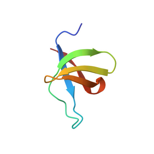
    The solution structure of the DNA binding domain of HIV-1 integrase (residues 220-270) has been determined by multidimensional NMR spectroscopy. The protein is a dimer in solution, and each subunit is composed of a five-stranded beta-barrel with a topology very similar to that of the SH3 domain. The dimer is formed by a stacked beta-interface comprising strands 2, 3, and 4, with the two triple-stranded antiparallel beta-sheets, one from each subunit, oriented antiparallel to each other. One surface of the dimer, bounded by the loop between strands beta 1 and beta 2, forms a saddle-shaped groove with dimensions of approximately 24 x 23 x 12 A in cross section. Lys264, which has been shown from mutational data to be involved in DNA binding, protrudes from this surface, implicating the saddle-shaped groove as the potential DNA binding site.


  • Organizational Affiliation
    • Laboratory of Chemical Physics, National Institute of Diabetes and Digestive and Kidney Diseases, National Institutes of Health, Bethesda, Maryland 20892-0520, USA.

Macromolecule Content 

  • Total Structure Weight: 12.28 kDa 
  • Atom Count: 866 
  • Modeled Residue Count: 104 
  • Deposited Residue Count: 104 
  • Unique protein chains: 1

Macromolecules

Find similar proteins by:|  3D Structure
Entity ID: 1
MoleculeChains  Sequence LengthOrganismDetailsImage
HIV-1 INTEGRASE
A, B
52Human immunodeficiency virus 1Mutation(s): 0 
UniProt
Find proteins for P04586 (Human immunodeficiency virus type 1 group M subtype D (isolate Z6))
Explore P04586 
Go to UniProtKB:  P04586
Entity Groups
Sequence Clusters30% Identity50% Identity70% Identity90% Identity95% Identity100% Identity
UniProt GroupP04586
Sequence Annotations
Expand
Reference Sequence

Experimental Data & Validation

Experimental Data

  • Method: SOLUTION NMR
  • Conformers Submitted: 40 

Structure Validation

View Full Validation Report


Entry History 

Deposition Data

Revision History  (Full details and data files)

  • Version 1.0: 1996-07-11
    Type: Initial release
  • Version 1.1: 2008-03-24
    Changes: Version format compliance
  • Version 1.2: 2011-07-13
    Changes: Version format compliance
  • Version 1.3: 2022-02-23
    Changes: Database references, Derived calculations, Other, Structure summary
  • Version 1.4: 2024-05-22
    Changes: Data collection