1IDE | pdb_00001ide

ISOCITRATE DEHYDROGENASE Y160F MUTANT STEADY-STATE INTERMEDIATE COMPLEX (LAUE DETERMINATION)


Experimental Data Snapshot

  • Method: X-RAY DIFFRACTION
  • Resolution: 2.50 Å
  • R-Value Work: 
    0.192 (Depositor) 
  • R-Value Observed: 
    0.192 (Depositor) 

wwPDB Validation 3D Report Full Report

Validation slider image for 1IDE

This is version 1.5 of the entry. See complete history

Literature

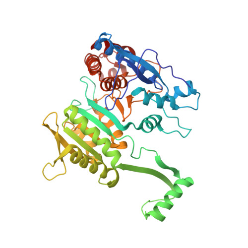
Mutagenesis and Laue structures of enzyme intermediates: isocitrate dehydrogenase.

Bolduc, J.M.Dyer, D.H.Scott, W.G.Singer, P.Sweet, R.M.Koshland Jr., D.E.Stoddard, B.L.

(1995) Science 268: 1312-1318

  • DOI: https://doi.org/10.1126/science.7761851
  • Primary Citation Related Structures: 
    1IDC, 1IDD, 1IDE, 1IDF

  • PubMed Abstract: 

    Site-directed mutagenesis and Laue diffraction data to 2.5 A resolution were used to solve the structures of two sequential intermediates formed during the catalytic actions of isocitrate dehydrogenase. Both intermediates are distinct from the enzyme-substrate and enzyme-product complexes. Mutation of key catalytic residues changed the rate determining steps so that protein and substrate intermediates within the overall reaction pathway could be visualized.


  • Organizational Affiliation
    • Fred Hutchinson Cancer Research Center, Program in Structural Biology, Seattle, WA 98104, USA.

Macromolecule Content 

  • Total Structure Weight: 46.75 kDa 
  • Atom Count: 3,259 
  • Modeled Residue Count: 414 
  • Deposited Residue Count: 416 
  • Unique protein chains: 1

Macromolecules

Find similar proteins by:|  3D Structure
Entity ID: 1
MoleculeChains  Sequence LengthOrganismDetailsImage
ISOCITRATE DEHYDROGENASE416Escherichia coliMutation(s): 1 
Gene Names: ICD
EC: 1.1.1.42
UniProt
Find proteins for P08200 (Escherichia coli (strain K12))
Explore P08200 
Go to UniProtKB:  P08200
Entity Groups
Sequence Clusters30% Identity50% Identity70% Identity90% Identity95% Identity100% Identity
UniProt GroupP08200
Sequence Annotations
Expand
Reference Sequence

Small Molecules

Ligands 3 Unique
IDChains Name / Formula / InChI Key2D Diagram3D Interactions
NAP

Query on NAP


Download:Ideal Coordinates CCD File
D [auth A]NADP NICOTINAMIDE-ADENINE-DINUCLEOTIDE PHOSPHATE
C21 H28 N7 O17 P3
XJLXINKUBYWONI-NNYOXOHSSA-N
ICT

Query on ICT


Download:Ideal Coordinates CCD File
C [auth A]ISOCITRIC ACID
C6 H8 O7
ODBLHEXUDAPZAU-ZAFYKAAXSA-N
MG

Query on MG


Download:Ideal Coordinates CCD File
B [auth A]MAGNESIUM ION
Mg
JLVVSXFLKOJNIY-UHFFFAOYSA-N

Experimental Data & Validation

Experimental Data

  • Method: X-RAY DIFFRACTION
  • Resolution: 2.50 Å
  • R-Value Work:  0.192 (Depositor) 
  • R-Value Observed: 0.192 (Depositor) 
Space Group: P 43 21 2
Unit Cell:
Length ( Å )Angle ( ˚ )
a = 105.1α = 90
b = 105.1β = 90
c = 150.3γ = 90
Software Package:
Software NamePurpose
X-PLORmodel building
X-PLORrefinement
OXFORDdata reduction
X-PLORphasing

Structure Validation

View Full Validation Report


Entry History 

Deposition Data

Revision History  (Full details and data files)

  • Version 1.0: 1996-03-08
    Type: Initial release
  • Version 1.1: 2008-03-03
    Changes: Version format compliance
  • Version 1.2: 2011-07-13
    Changes: Derived calculations, Version format compliance
  • Version 1.3: 2017-11-29
    Changes: Derived calculations, Other
  • Version 1.4: 2021-11-03
    Changes: Database references, Derived calculations
  • Version 1.5: 2024-02-07
    Changes: Data collection