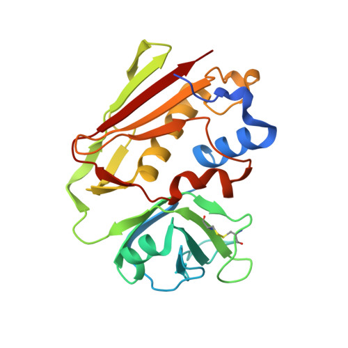
1I4G

Crystal structure of Staphylococcal enterotoxin A mutant H187A with reduced Zn2+ affinity


Experimental Data Snapshot

  • Method: X-RAY DIFFRACTION
  • Resolution: 2.10 Å
  • R-Value Free: 0.239 
  • R-Value Work: 0.178 

Starting Model: experimental
View more details

wwPDB Validation   3D Report Full Report


This is version 1.6 of the entry. See complete history


Literature

Cooperative zinc binding in a staphylococcal enterotoxin A mutant mimics the SEA-MHC class II interaction

Hakansson, M.Antonsson, P.Bjork, P.Svensson, L.A.

(2001) J Biol Inorg Chem 6: 757-762

  • DOI: https://doi.org/10.1007/s007750100251
  • Primary Citation of Related Structures:  
    1I4G, 1I4H

  • PubMed Abstract: 

    The structure of a mutant form of staphylococcal enterotoxin A (SEA) has been determined to 2.1 A resolution. The studied SEA substitution H187-->A187 (SEAH187A) leads to an almost 10-fold reduction of the binding to major histocompatibility complex (MHC) class II. H187 is important for this interaction since it coordinates Zn2+. The zinc ion is thought to hold MHC class II and SEA together in a complex. Interestingly, only one of two molecules in the asymmetric unit binds Zn2+. H225, D227, a water molecule, and H44 from a symmetry-related molecule ligate Zn2+. The symmetry-related histidine is necessary for this substituted Zn2+ site to bind to Zn2+ at low zinc concentration (no Zn2+ added). Since a water molecule replaces the missing H187, H44 binds Zn2+ at the position where betaH81 from MHC class II probably will bind. Dynamic light scattering analysis reveals that in solution as well as in the crystal lattice the SEA(H187A) mutant forms aggregates. The substitution per se does not cause aggregation since wild-type SEA also forms aggregates. Addition of EDTA reduces the size of the aggregates, indicating a cross-linking function of Zn2+. In agreement with the biological function, the aggregation is weak (i.e. not revealed by gel filtration) and non-specific.


  • Organizational Affiliation

    Department of Molecular Biophysics, Center for Chemistry and Chemical Engineering, P.O. Box 124, 221 00 Lund, Sweden. maria.hakansson@mbfys.lu.se


Macromolecules
Find similar proteins by:  (by identity cutoff)  |  3D Structure
Entity ID: 1
MoleculeChains Sequence LengthOrganismDetailsImage
ENTEROTOXIN TYPE A
A, B
233Staphylococcus aureusMutation(s): 1 
UniProt
Find proteins for P0A0L2 (Staphylococcus aureus)
Explore P0A0L2 
Go to UniProtKB:  P0A0L2
Entity Groups  
Sequence Clusters30% Identity50% Identity70% Identity90% Identity95% Identity100% Identity
UniProt GroupP0A0L2
Sequence Annotations
Expand
  • Reference Sequence
Small Molecules
Ligands 2 Unique
IDChains Name / Formula / InChI Key2D Diagram3D Interactions
SO4
Query on SO4

Download Ideal Coordinates CCD File 
D [auth B]SULFATE ION
O4 S
QAOWNCQODCNURD-UHFFFAOYSA-L
ZN
Query on ZN

Download Ideal Coordinates CCD File 
C [auth A]ZINC ION
Zn
PTFCDOFLOPIGGS-UHFFFAOYSA-N
Experimental Data & Validation

Experimental Data

  • Method: X-RAY DIFFRACTION
  • Resolution: 2.10 Å
  • R-Value Free: 0.239 
  • R-Value Work: 0.178 
  • Space Group: P 21 21 21
Unit Cell:
Length ( Å )Angle ( ˚ )
a = 73.66α = 90
b = 84.83β = 90
c = 86.35γ = 90
Software Package:
Software NamePurpose
XDSdata scaling
AUTOMARdata reduction
AMoREphasing
X-PLORrefinement
XDSdata reduction

Structure Validation

View Full Validation Report


Entry History 

Deposition Data

Revision History  (Full details and data files)

  • Version 1.0: 2001-02-28
    Type: Initial release
  • Version 1.1: 2008-04-27
    Changes: Version format compliance
  • Version 1.2: 2011-07-13
    Changes: Version format compliance
  • Version 1.3: 2017-10-04
    Changes: Refinement description
  • Version 1.4: 2021-10-27
    Changes: Database references, Derived calculations
  • Version 1.5: 2023-08-09
    Changes: Data collection, Refinement description
  • Version 1.6: 2024-10-16
    Changes: Structure summary