1I25 | pdb_00001i25

Three dimensional solution structure of huwentoxin-II by 2D 1H-NMR


Experimental Data Snapshot

  • Method: SOLUTION NMR
  • Conformers Calculated: 50 
  • Conformers Submitted: 10 
  • Selection Criteria: structures with the lowest energy 

wwPDB Validation 3D Report Full Report

Validation slider image for 1I25

This is version 1.4 of the entry. See complete history

Literature

The structure of spider toxin huwentoxin-II with unique disulfide linkage: evidence for structural evolution.

Shu, Q.Lu, S.Y.Gu, X.C.Liang, S.P.

(2002) Protein Sci 11: 245-252

  • DOI: https://doi.org/10.1110/ps.30502
  • Primary Citation Related Structures: 
    1I25

  • PubMed Abstract: 

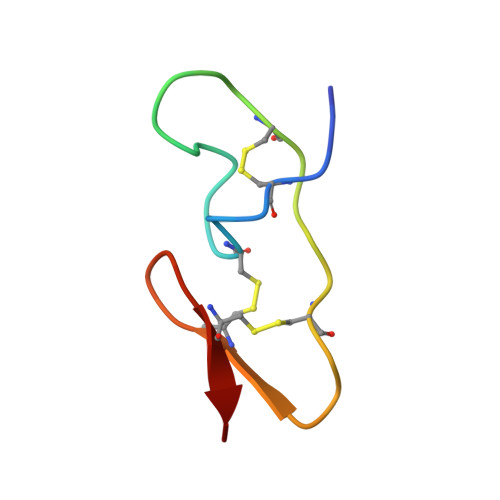
    The three-dimensional structure of huwentoxin-II (HWTX-II), an insecticidal peptide purified from the venom of spider Selenocosmia huwena with a unique disulfide bond linkage as I-III, II-V, and IV-VI, has been determined using 2D (1)H-NMR. The resulting structure of HWTX-II contains two beta-turns (C4-S7 and K24-W27) and a double-stranded antiparallel beta-sheet (W27-C29 and C34-K36). Although the C-terminal double-stranded beta-sheet cross-linked by two disulfide bonds (II-V and IV-VI in HWTX-II, II-V and III-VI in the ICK molecules) is conserved both in HWTX-II and the ICK molecules, the structure of HWTX-II is unexpected absence of the cystine knot because of its unique disulfide linkage. It suggests that HWTX-II adopts a novel scaffold different from the ICK motif that is adopted by all other spider toxin structures elucidated thus far. Furthermore, the structure of HWTX-II, which conforms to the disulfide-directed beta-hairpin (DDH) motif, not only supports the hypothesis that the ICK is a minor elaboration of the more ancestral DDH motif but also suggests that HWTX-II may have evolved from the same structural ancestor.


  • Organizational Affiliation
    • Life Science College, Peking University, Beijing 100871, People's Republic of China.

Macromolecule Content 

  • Total Structure Weight: 4.3 kDa 
  • Atom Count: 295 
  • Modeled Residue Count: 37 
  • Deposited Residue Count: 37 
  • Unique protein chains: 1

Macromolecules

Find similar proteins by:|  3D Structure
Entity ID: 1
MoleculeChains  Sequence LengthOrganismDetailsImage
HUWENTOXIN-II37Cyriopagopus schmidtiMutation(s): 0 
Membrane Entity: Yes 
UniProt
Find proteins for P82959 (Cyriopagopus schmidti)
Explore P82959 
Go to UniProtKB:  P82959
Entity Groups
Sequence Clusters30% Identity50% Identity70% Identity90% Identity95% Identity100% Identity
UniProt GroupP82959
Sequence Annotations
Expand
Reference Sequence

Experimental Data & Validation

Experimental Data

  • Method: SOLUTION NMR
  • Conformers Calculated: 50 
  • Conformers Submitted: 10 
  • Selection Criteria: structures with the lowest energy 

Structure Validation

View Full Validation Report


Entry History 

Deposition Data

Revision History  (Full details and data files)

  • Version 1.0: 2001-02-14
    Type: Initial release
  • Version 1.1: 2008-04-27
    Changes: Version format compliance
  • Version 1.2: 2011-07-13
    Changes: Version format compliance
  • Version 1.3: 2017-11-29
    Changes: Derived calculations
  • Version 1.4: 2024-11-06
    Changes: Data collection, Database references, Structure summary