Solution structure of the catalytic domain of gammadelta resolvase. Implications for the mechanism of catalysis.
Pan, B., Maciejewski, M.W., Marintchev, A., Mullen, G.P.(2001) J Mol Biol 310: 1089-1107
- PubMed: 11501998 Search on PubMed
- DOI: https://doi.org/10.1006/jmbi.2001.4821
- Primary Citation Related Structures:
1GHT, 1HX7 - PubMed Abstract:
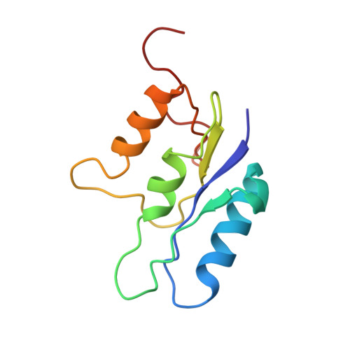
The site-specific DNA recombinase, gammadelta resolvase, from Escherichia coli catalyzes recombination of res site-containing plasmid DNA to two catenated circular DNA products. The catalytic domain (residues 1-105), lacking a C-terminal dimerization interface, has been constructed and the NMR solution structure of the monomer determined. The RMSD of the NMR conformers for residues 2-92 excluding residues 37-45 and 64-73 is 0.41 A for backbone atoms and 0.88 A for all heavy atoms. The NMR solution structure of the monomeric catalytic domain (residues 1-105) was found to be formed by a four-stranded parallel beta-sheet surrounded by three helices. The catalytic domain (residues 1-105), deficient in the C-terminal dimerization domain, was monomeric at high salt concentration, but displayed unexpected dimerization at lower ionic strength. The unique solution dimerization interface at low ionic strength was mapped by NMR. With respect to previous crystal structures of the dimeric catalytic domain (residues 1-140), differences in the average conformation of active-site residues were found at loop 1 containing the catalytic S10 nucleophile, the beta1 strand containing R8, and at loop 3 containing D67, R68 and R71, which are required for catalysis. The active-site loops display high-frequency and conformational backbone dynamics and are less well defined than the secondary structures. In the solution structure, the D67 side-chain is proximal to the S10 side-chain making the D67 carboxylate group a candidate for activation of S10 through general base catalysis. Four conserved Arg residues can function in the activation of the phosphodiester for nucleophilic attack by the S10 hydroxyl group. A mechanism for covalent catalysis by this class of recombinases is proposed that may be related to dimer interface dissociation.
- Department of Biochemistry, University of Connecticut Health Center, Farmington 06032, USA.
Organizational Affiliation: