1HVB | pdb_00001hvb

CRYSTAL STRUCTURE OF STREPTOMYCES R61 DD-PEPTIDASE COMPLEXED WITH A NOVEL CEPHALOSPORIN ANALOG OF CELL WALL PEPTIDOGLYCAN


Experimental Data Snapshot

  • Method: X-RAY DIFFRACTION
  • Resolution: 1.17 Å
  • R-Value Free: 
    0.153 (Depositor) 
  • R-Value Work: 
    0.113 (Depositor) 
  • R-Value Observed: 
    0.114 (Depositor) 

Starting Model: experimental
View more details

wwPDB Validation 3D Report Full Report

Validation slider image for 1HVB

This is version 1.5 of the entry. See complete history. 

Literature

A 1.2-A snapshot of the final step of bacterial cell wall biosynthesis.

Lee, W., McDonough, M.A., Kotra, L., Li, Z.H., Silvaggi, N.R., Takeda, Y., Kelly, J.A., Mobashery, S.

(2001) Proc Natl Acad Sci U S A 98: 1427-1431

  • DOI: https://doi.org/10.1073/pnas.98.4.1427
  • Primary Citation Related Structures: 
    1HVB

  • PubMed Abstract: 

    The cell wall imparts structural strength and shape to bacteria. It is made up of polymeric glycan chains with peptide branches that are cross-linked to form the cell wall. The cross-linking reaction, catalyzed by transpeptidases, is the last step in cell wall biosynthesis. These enzymes are members of the family of penicillin-binding proteins, the targets of beta-lactam antibiotics. We report herein the structure of a penicillin-binding protein complexed with a cephalosporin designed to probe the mechanism of the cross-linking reaction catalyzed by transpeptidases. The 1.2-A resolution x-ray structure of this cephalosporin bound to the active site of the bifunctional serine type D-alanyl-D-alanine carboxypeptidase/transpeptidase (EC ) from Streptomyces sp. strain R61 reveals how the two peptide strands from the polymeric substrates are sequestered in the active site of a transpeptidase. The structure of this complex provides a snapshot of the enzyme and the bound cell wall components poised for the final and critical cross-linking step of cell wall biosynthesis.


  • Organizational Affiliation: 
    • Department of Molecular and Cell Biology, University of Connecticut, Storrs, CT 06269-3125, USA.

Macromolecule Content 

  • Total Structure Weight: 38.17 kDa 
  • Atom Count: 3,318 
  • Modeled Residue Count: 346 
  • Deposited Residue Count: 349 
  • Unique protein chains: 1

Macromolecules

Find similar proteins by:|  3D Structure
Entity ID: 1
MoleculeChains  Sequence LengthOrganismDetailsImage
D-ALANYL-D-ALANINE CARBOXYPEPTIDASE349Streptomyces sp. R61Mutation(s): 0 
EC: 3.4.16.4
UniProt
Find proteins for P15555 (Streptomyces sp. (strain R61))
Explore P15555 
Go to UniProtKB:  P15555
Entity Groups
Sequence Clusters30% Identity50% Identity70% Identity90% Identity95% Identity100% Identity
UniProt GroupP15555
Sequence Annotations
Expand
Reference Sequence

Small Molecules

Ligands 1 Unique
IDChains Name / Formula / InChI Key2D Diagram3D Interactions
CEH

Query on CEH


Download:Ideal Coordinates CCD File
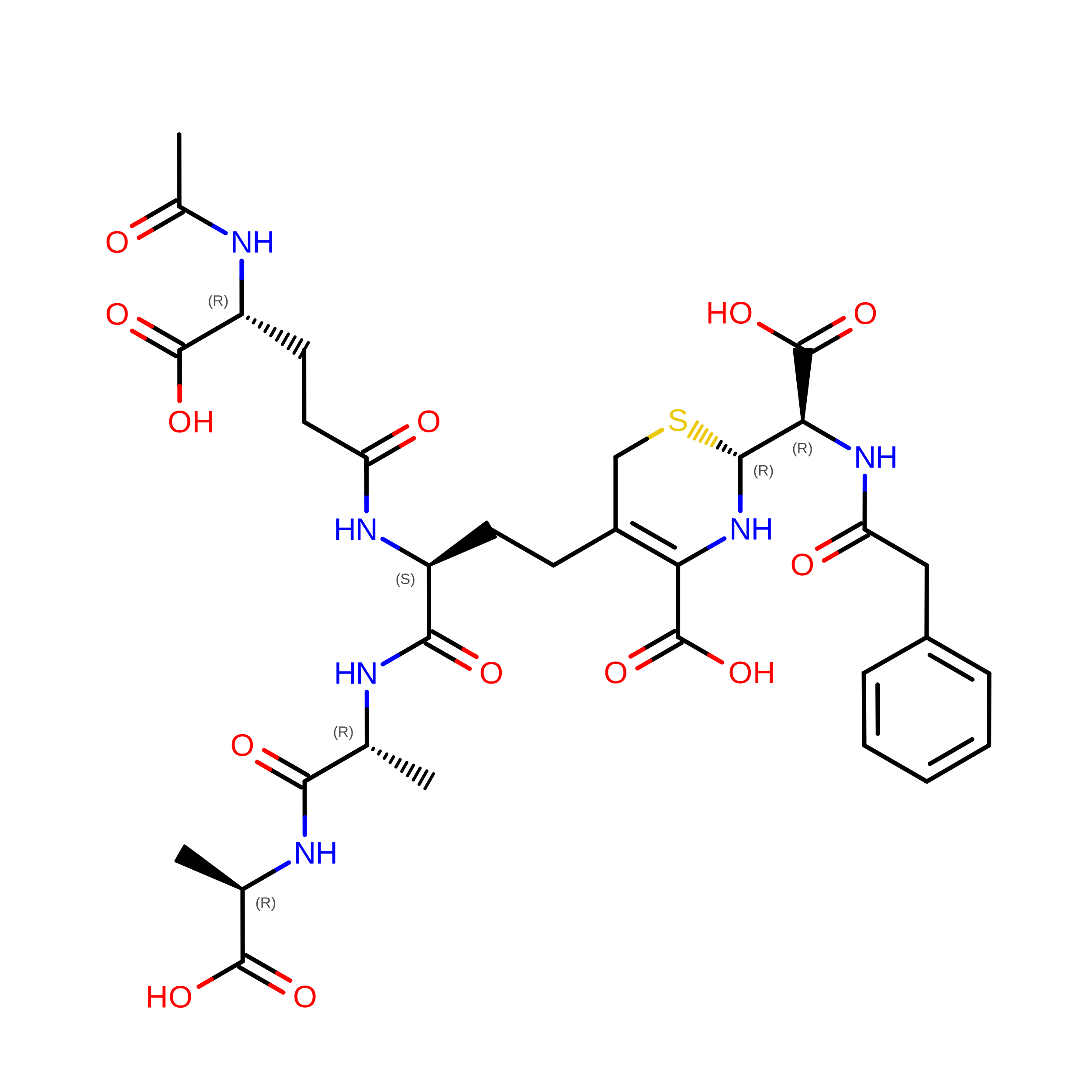
B [auth A]5-{3-(S)-(4-(R)-ACETYLAMINO-4-CARBOXY-BUTYRYLAMINO)-3-[1-(R)-(1-(R)-CARBOXY-ETHYLCARBAMOYL)-ETHYLCARBAMOYL]-PROPYL}-2-( CARBOXY-PHENYLACETYLAMINO-METHYL)-3,6-DIHYDRO-2H-[1,3]THIAZINE-4-CARBOXYLIC ACID
C32 H42 N6 O13 S
PEUIVMLYMKXUBF-PULJXETJSA-N

Experimental Data & Validation

Experimental Data

  • Method: X-RAY DIFFRACTION
  • Resolution: 1.17 Å
  • R-Value Free:  0.153 (Depositor) 
  • R-Value Work:  0.113 (Depositor) 
  • R-Value Observed: 0.114 (Depositor) 
Space Group: P 21 21 21
Unit Cell:
Length ( Å )Angle ( ˚ )
a = 50.778α = 90
b = 66.664β = 90
c = 100.928γ = 90
Software Package:
Software NamePurpose
SHELXL-97refinement
SCALEPACKdata scaling
CNSrefinement
DENZOdata reduction
CNSphasing

Structure Validation

View Full Validation Report


Entry History 

Deposition Data

Revision History  (Full details and data files)

  • Version 1.0: 2001-02-07
    Type: Initial release
  • Version 1.1: 2008-04-27
    Changes: Version format compliance
  • Version 1.2: 2011-07-13
    Changes: Source and taxonomy, Version format compliance
  • Version 1.3: 2019-07-24
    Changes: Data collection, Derived calculations, Refinement description
  • Version 1.4: 2023-08-09
    Changes: Data collection, Database references, Derived calculations, Refinement description, Structure summary
  • Version 1.5: 2024-10-30
    Changes: Structure summary