An extended hydrophobic core induces EF-hand swapping.
Hakansson, M., Svensson, A., Fast, J., Linse, S.(2001) Protein Sci 10: 927-933
- PubMed: 11316872 Search on PubMedSearch on PubMed Central
- DOI: https://doi.org/10.1110/ps.47501
- Primary Citation Related Structures:
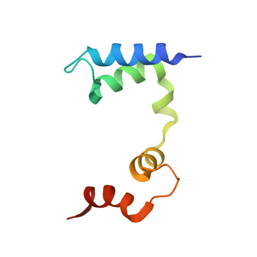
1HT9 - PubMed Abstract:
The structure of calbindin D(9k) with two substitutions was determined by X-ray crystallography at 1.8-A resolution. Unlike wild-type calbindin D(9k), which is a monomeric protein with two EF-hands, the structure of the mutated calbindin D(9k) reveals an intertwined dimer. In the dimer, two EF-hands of the monomers have exchanged places, and thus a 3D domain-swapped dimer has been formed. EF-hand I of molecule A is packed toward EF-hand II of molecule B and vice versa. The formation of a hydrophobic cluster, in a region linking the EF-hands, promotes the conversion of monomers to 3D domain-swapped dimers. We propose a mechanism by which domain swapping takes place via the apo form of calbindin D(9k). Once formed, the calbindin D(9k) dimers are remarkably stable, as with even larger misfolded aggregates like amyloids. Thus calbindin D(9k) dimers cannot be converted to monomers by dilution. However, heating can be used for conversion, indicating high energy barriers separating monomers from dimers.
- Molecular Biophysics, Center for Chemistry and Chemical Engineering, Lund University, S-221 00 Lund, Sweden. Maria.Hakansson@mbfys.lu.se
Organizational Affiliation: