1HRB | pdb_00001hrb

ATOMIC MODELS FOR THE POLYPEPTIDE BACKBONES OF MYOHEMERYTHRIN AND HEMERYTHRIN


Experimental Data Snapshot

  • Method: X-RAY DIFFRACTION
  • Resolution: 5.50 Å

wwPDB Validation 3D Report Full Report

Validation slider image for 1HRB

This is version 1.3 of the entry. See complete history

Literature

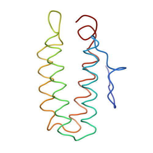
Atomic models for the polypeptide backbones of myohemerythrin and hemerythrin.

Hendrickson, W.A.Ward, K.B.

(1975) Biochem Biophys Res Commun 66: 1349-1356

Macromolecule Content 

  • Total Structure Weight: 27.02 kDa 
  • Atom Count: 230 
  • Modeled Residue Count: 226 
  • Deposited Residue Count: 226 
  • Unique protein chains: 1

Macromolecules

Find similar proteins by:|  3D Structure
Entity ID: 1
MoleculeChains  Sequence LengthOrganismDetailsImage
HEMERYTHRIN B
A, B
113Phascolopsis gouldiiMutation(s): 0 
UniProt
Find proteins for P02244 (Phascolopsis gouldii)
Explore P02244 
Go to UniProtKB:  P02244
Entity Groups
Sequence Clusters30% Identity50% Identity70% Identity90% Identity95% Identity100% Identity
UniProt GroupP02244
Sequence Annotations
Expand
Reference Sequence

Experimental Data & Validation

Experimental Data

  • Method: X-RAY DIFFRACTION
  • Resolution: 5.50 Å
Space Group: P 4 2 2
Unit Cell:
Length ( Å )Angle ( ˚ )
a = 104.82α = 90
b = 104.82β = 90
c = 54.08γ = 90

Structure Validation

View Full Validation Report


Entry History 

Deposition Data

Revision History  (Full details and data files)

  • Version 1.0: 1978-09-28
    Type: Initial release
  • Version 1.1: 2008-03-24
    Changes: Version format compliance
  • Version 1.2: 2011-07-13
    Changes: Version format compliance
  • Version 1.3: 2024-02-07
    Changes: Data collection, Database references, Derived calculations, Other