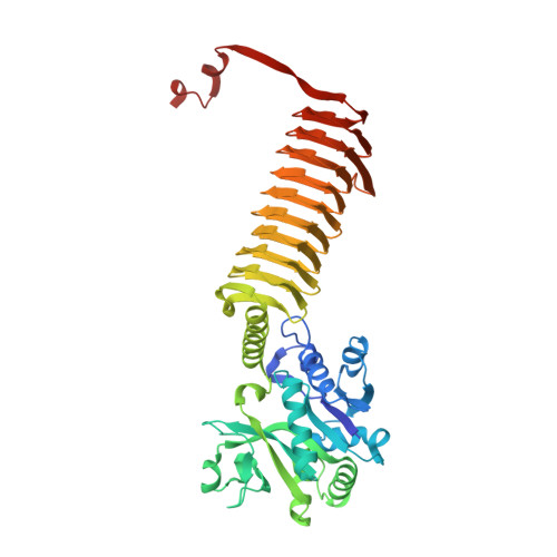
1HM8

CRYSTAL STRUCTURE OF S.PNEUMONIAE N-ACETYLGLUCOSAMINE-1-PHOSPHATE URIDYLTRANSFERASE, GLMU, BOUND TO ACETYL COENZYME A


Experimental Data Snapshot

  • Method: X-RAY DIFFRACTION
  • Resolution: 2.50 Å
  • R-Value Free: 0.243 
  • R-Value Work: 0.184 
  • R-Value Observed: 0.184 

wwPDB Validation   3D Report Full Report


Ligand Structure Quality Assessment 

Created with Raphaël 2.3.0Worse 01 BetterLigand structure goodness of fit to experimental dataBest fitted ACOClick on this verticalbar to view details

This is version 1.3 of the entry. See complete history


Literature

Crystal structure of Streptococcus pneumoniae N-acetylglucosamine-1-phosphate uridyltransferase bound to acetyl-coenzyme A reveals a novel active site architecture.

Sulzenbacher, G.Gal, L.Peneff, C.Fassy, F.Bourne, Y.

(2001) J Biol Chem 276: 11844-11851

  • DOI: https://doi.org/10.1074/jbc.M011225200
  • Primary Citation of Related Structures:  
    1HM0, 1HM8, 1HM9

  • PubMed Abstract: 

    The bifunctional bacterial enzyme N-acetyl-glucosamine-1-phosphate uridyltransferase (GlmU) catalyzes the two-step formation of UDP-GlcNAc, a fundamental precursor in bacterial cell wall biosynthesis. With the emergence of new resistance mechanisms against beta-lactam and glycopeptide antibiotics, the biosynthetic pathway of UDP-GlcNAc represents an attractive target for drug design of new antibacterial agents. The crystal structures of Streptococcus pneumoniae GlmU in unbound form, in complex with acetyl-coenzyme A (AcCoA) and in complex with both AcCoA and the end product UDP-GlcNAc, have been determined and refined to 2.3, 2.5, and 1.75 A, respectively. The S. pneumoniae GlmU molecule is organized in two separate domains connected via a long alpha-helical linker and associates as a trimer, with the 50-A-long left-handed beta-helix (LbetaH) C-terminal domains packed against each other in a parallel fashion and the C-terminal region extended far away from the LbetaH core and exchanged with the beta-helix from a neighboring subunit in the trimer. AcCoA binding induces the formation of a long and narrow tunnel, enclosed between two adjacent LbetaH domains and the interchanged C-terminal region of the third subunit, giving rise to an original active site architecture at the junction of three subunits.


  • Organizational Affiliation

    AFMB-UMR6098, 31 Chemin Joseph Aiguier, 13402 Marseille Cedex 20, France.


Macromolecules
Find similar proteins by:  (by identity cutoff)  |  3D Structure
Entity ID: 1
MoleculeChains Sequence LengthOrganismDetailsImage
UDP-N-ACETYLGLUCOSAMINE-1-PHOSPHATE URIDYLTRANSFERASE
A, B
468Streptococcus pneumoniaeMutation(s): 0 
Gene Names: GLMU
EC: 2.7.7.23 (PDB Primary Data), 2.3.1.157 (UniProt)
UniProt
Find proteins for Q97R46 (Streptococcus pneumoniae serotype 4 (strain ATCC BAA-334 / TIGR4))
Explore Q97R46 
Go to UniProtKB:  Q97R46
Entity Groups  
Sequence Clusters30% Identity50% Identity70% Identity90% Identity95% Identity100% Identity
UniProt GroupQ97R46
Sequence Annotations
Expand
  • Reference Sequence
Experimental Data & Validation

Experimental Data

  • Method: X-RAY DIFFRACTION
  • Resolution: 2.50 Å
  • R-Value Free: 0.243 
  • R-Value Work: 0.184 
  • R-Value Observed: 0.184 
  • Space Group: H 3
Unit Cell:
Length ( Å )Angle ( ˚ )
a = 92.459α = 90
b = 92.459β = 90
c = 279.562γ = 120
Software Package:
Software NamePurpose
CNSrefinement
DENZOdata reduction
CCP4data scaling
CNSphasing

Structure Validation

View Full Validation Report


Ligand Structure Quality Assessment 

Created with Raphaël 2.3.0Worse 01 BetterLigand structure goodness of fit to experimental dataBest fitted ACOClick on this verticalbar to view details

Entry History 

Deposition Data

Revision History  (Full details and data files)

  • Version 1.0: 2001-11-30
    Type: Initial release
  • Version 1.1: 2008-04-27
    Changes: Version format compliance
  • Version 1.2: 2011-07-13
    Changes: Derived calculations, Version format compliance
  • Version 1.3: 2024-02-07
    Changes: Data collection, Database references, Derived calculations