1HIN

STRUCTURAL EVIDENCE FOR INDUCED FIT AS A MECHANISM FOR ANTIBODY-ANTIGEN RECOGNITION


Experimental Data Snapshot

  • Method: X-RAY DIFFRACTION
  • Resolution: 3.10 Å
  • R-Value Work: 0.220 
  • R-Value Observed: 0.220 

wwPDB Validation   3D Report Full Report


This is version 1.3 of the entry. See complete history


Literature

Structural evidence for induced fit as a mechanism for antibody-antigen recognition.

Rini, J.M.Schulze-Gahmen, U.Wilson, I.A.

(1992) Science 255: 959-965

  • DOI: https://doi.org/10.1126/science.1546293
  • Primary Citation of Related Structures:  
    1HIL, 1HIM, 1HIN

  • PubMed Abstract: 

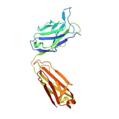
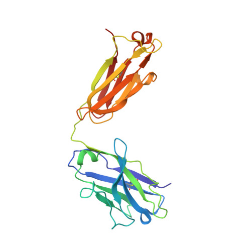
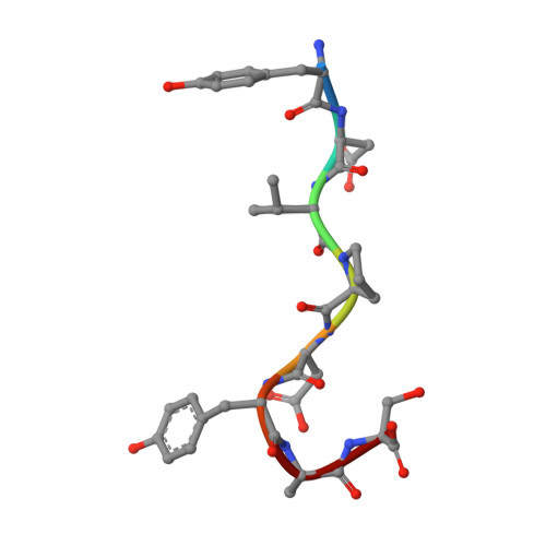
    The three-dimensional structure of a specific antibody (Fab 17/9) to a peptide immunogen from influenza virus hemagglutinin [HA1(75-110)] and two independent crystal complexes of this antibody with bound peptide (TyrP100-LeuP108) have been determined by x-ray crystallographic techniques at 2.0 A, 2.9 A, and 3.1 A resolution, respectively. The nonapeptide antigen assumes a type I beta turn in the antibody combining site and interacts primarily with the Fab hypervariable loops L3, H2, and H3. Comparison of the bound and unbound Fab structures shows that a major rearrangement in the H3 loop accompanies antigen binding. This conformational change results in the creation of a binding pocket for the beta turn of the peptide, allowing TyrP105 to be accommodated. The conformation of the peptide bound to the antibody shows similarity to its cognate sequence in the HA1, suggesting a possible mechanism for the cross-reactivity of this Fab with monomeric hemagglutinin. The structures of the free and antigen bound antibodies demonstrate the flexibility of the antibody combining site and provide an example of induced fit as a mechanism for antibody-antigen recognition.


  • Organizational Affiliation

    Department of Molecular Biology, Scripps Research Institute, La Jolla, CA 92037.


Macromolecules
Find similar proteins by:  (by identity cutoff)  |  3D Structure
Entity ID: 1
MoleculeChains Sequence LengthOrganismDetailsImage
IGG2A-KAPPA 17/9 FAB (LIGHT CHAIN)A [auth L]217Mus musculusMutation(s): 0 
Entity Groups  
Sequence Clusters30% Identity50% Identity70% Identity90% Identity95% Identity100% Identity
Sequence Annotations
Expand
  • Reference Sequence
Find similar proteins by:  (by identity cutoff)  |  3D Structure
Entity ID: 2
MoleculeChains Sequence LengthOrganismDetailsImage
IGG2A-KAPPA 17/9 FAB (HEAVY CHAIN)B [auth H]220Mus musculusMutation(s): 0 
Entity Groups  
Sequence Clusters30% Identity50% Identity70% Identity90% Identity95% Identity100% Identity
Sequence Annotations
Expand
  • Reference Sequence

Find similar proteins by:  Sequence   |   3D Structure  

Entity ID: 3
MoleculeChains Sequence LengthOrganismDetailsImage
INFLUENZA HEMAGGLUTININ HA1 (STRAIN X47) (RESIDUES 100-107)C [auth P]8N/AMutation(s): 0 
Sequence Annotations
Expand
  • Reference Sequence
Experimental Data & Validation

Experimental Data

  • Method: X-RAY DIFFRACTION
  • Resolution: 3.10 Å
  • R-Value Work: 0.220 
  • R-Value Observed: 0.220 
  • Space Group: P 1 21 1
Unit Cell:
Length ( Å )Angle ( ˚ )
a = 63.5α = 90
b = 73.4β = 117.1
c = 62.7γ = 90
Software Package:
Software NamePurpose
X-PLORmodel building
X-PLORrefinement
X-PLORphasing

Structure Validation

View Full Validation Report


Entry History 

Deposition Data

Revision History  (Full details and data files)

  • Version 1.0: 1994-01-31
    Type: Initial release
  • Version 1.1: 2008-03-24
    Changes: Version format compliance
  • Version 1.2: 2011-07-13
    Changes: Version format compliance
  • Version 1.3: 2024-06-05
    Changes: Data collection, Database references, Derived calculations, Other