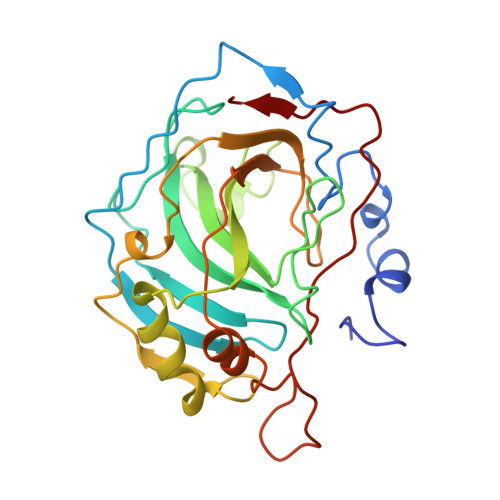
1HEA

CARBONIC ANHYDRASE II (CARBONATE DEHYDRATASE) (HCA II) (E.C.4.2.1.1) MUTANT WITH LEU 198 REPLACED BY ARG (L198R)


Experimental Data Snapshot

  • Method: X-RAY DIFFRACTION
  • Resolution: 2.00 Å
  • R-Value Observed: 0.182 

wwPDB Validation   3D Report Full Report


This is version 1.4 of the entry. See complete history


Literature

Structural consequences of hydrophilic amino acid substitutions in the hydrophobic pocket of human carbonic anhydrase II.

Nair, S.K.Christianson, D.W.

(1993) Biochemistry 32: 4506-4514

  • DOI: https://doi.org/10.1021/bi00068a005
  • Primary Citation of Related Structures:  
    1HEA, 1HEB, 1HEC, 1HED

  • PubMed Abstract: 

    The three-dimensional structures of Leu-198-->Glu, Leu-198-->His, Leu-198-->Arg, and Leu-198-->Ala variants of human carbonic anhydrase II (CAII) have each been determined by X-ray crystallographic methods to a resolution of 2.0 A. The side chain of Leu-198 is located at the mouth of the active site hydrophobic pocket, and this pocket is required for substrate association. Hydrophobic-->hydrophilic amino acid substitutions at the mouth of the pocket decrease kcat/KM for CO2 hydration: the CO2 hydrase activities of Leu-198-->Glu, Leu-198-->His, and Leu-198-->Arg CAIIs are diminished 19-fold, 10-fold, and 17-fold, respectively, relative to the wild-type enzyme; however, the substitution of a compact aliphatic side chain for Leu-198 has a smaller effect on catalysis, in that Leu-198-->Ala CAII exhibits only a 3-fold decrease in CO2 hydrase activity [Krebs, J. F., Rana, F., Dluhy, R. A., & Fierke, C. A. (1993) Biochemistry (preceding paper in this issue)]. It is intriguing that CO2 hydrase activity is not severely diminished in Leu-198-->Arg CAII, even though the side chain of Arg-198 blocks the hydrophobic pocket. Therefore, the bulky side chain of Arg-198 must be reasonably mobile in order to accommodate substrate association. Significantly, a residue larger than the wild-type Leu-198 side chain does not necessarily block the substrate association pocket; e.g., the side chain of Glu-198 packs against a hydrophobic patch, the net result of which is a wider mouth for the pocket.(ABSTRACT TRUNCATED AT 250 WORDS)


  • Organizational Affiliation

    Department of Chemistry, University of Pennsylvania, Philadelphia 19104-6323.


Macromolecules
Find similar proteins by:  (by identity cutoff)  |  3D Structure
Entity ID: 1
MoleculeChains Sequence LengthOrganismDetailsImage
CARBONIC ANHYDRASE II260Homo sapiensMutation(s): 0 
EC: 4.2.1.1 (PDB Primary Data), 4.2.1.69 (UniProt)
UniProt & NIH Common Fund Data Resources
Find proteins for P00918 (Homo sapiens)
Explore P00918 
Go to UniProtKB:  P00918
PHAROS:  P00918
GTEx:  ENSG00000104267 
Entity Groups  
Sequence Clusters30% Identity50% Identity70% Identity90% Identity95% Identity100% Identity
UniProt GroupP00918
Sequence Annotations
Expand
  • Reference Sequence
Small Molecules
Ligands 2 Unique
IDChains Name / Formula / InChI Key2D Diagram3D Interactions
HG
Query on HG

Download Ideal Coordinates CCD File 
C [auth A]MERCURY (II) ION
Hg
BQPIGGFYSBELGY-UHFFFAOYSA-N
ZN
Query on ZN

Download Ideal Coordinates CCD File 
B [auth A]ZINC ION
Zn
PTFCDOFLOPIGGS-UHFFFAOYSA-N
Experimental Data & Validation

Experimental Data

  • Method: X-RAY DIFFRACTION
  • Resolution: 2.00 Å
  • R-Value Observed: 0.182 
  • Space Group: P 1 21 1
Unit Cell:
Length ( Å )Angle ( ˚ )
a = 42.7α = 90
b = 41.7β = 104.6
c = 73γ = 90
Software Package:
Software NamePurpose
PROLSQrefinement

Structure Validation

View Full Validation Report


Entry History 

Deposition Data

Revision History  (Full details and data files)

  • Version 1.0: 1992-10-15
    Type: Initial release
  • Version 1.1: 2008-03-24
    Changes: Version format compliance
  • Version 1.2: 2011-07-13
    Changes: Version format compliance
  • Version 1.3: 2017-11-29
    Changes: Derived calculations, Other
  • Version 1.4: 2024-02-07
    Changes: Data collection, Database references, Derived calculations