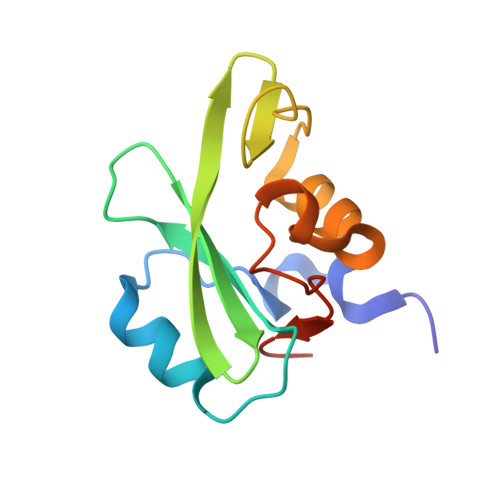
1H9O

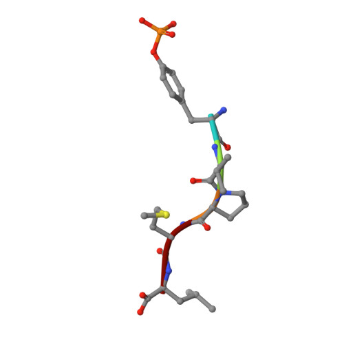
PHOSPHATIDYLINOSITOL 3-KINASE, P85-ALPHA SUBUNIT: C-TERMINAL SH2 DOMAIN COMPLEXED WITH A TYR751 PHOSPHOPEPTIDE FROM THE PDGF RECEPTOR, CRYSTAL STRUCTURE AT 1.79 A


Experimental Data Snapshot

  • Method: X-RAY DIFFRACTION
  • Resolution: 1.79 Å
  • R-Value Free: 0.219 
  • R-Value Work: 0.163 
  • R-Value Observed: 0.168 

Starting Model: experimental
View more details

wwPDB Validation   3D Report Full Report


This is version 1.5 of the entry. See complete history


Literature

NMR Trial Models: Experiences with the Colicin Immunity Protein Im7 and the P85Alpha C-Terminal Sh2-Peptide Complex

Pauptit, R.A.Dennis, C.A.Derbyshire, D.J.Breeze, A.L.Weston, S.A.Rowsell, S.Murshudov, G.N.

(2001) Acta Crystallogr D Biol Crystallogr 57: 1397

  • DOI: https://doi.org/10.1107/s0907444901012434
  • Primary Citation of Related Structures:  
    1H9O

  • PubMed Abstract: 

    Two cases of successful molecular replacement using NMR trial models are presented. One is the crystal structure of the Escherichia coli colicin immunity protein Im7; the other is a heretofore unreported crystal structure of a specific PDGF receptor-derived peptide complex of the carboxy-terminal SH2 domain from the p85alpha subunit of human phosphatidylinositol 3-OH kinase. In both cases, molecular replacement was non-trivial. Success was achieved using trial models that consisted of an ensemble of NMR structures from which the more flexible portions had been excised. Use of maximum-likelihood refinement proved critical to be able to refine the poor starting models. The challenges typical of the use of NMR trial models in molecular replacement are discussed.


  • Organizational Affiliation

    AstraZeneca, Mereside, Alderley Park, Macclesfield, Cheshire SK10 4TG, England. richard.pauptit@astrazeneca.com


Macromolecules
Find similar proteins by:  (by identity cutoff)  |  3D Structure
Entity ID: 1
MoleculeChains Sequence LengthOrganismDetailsImage
PHOSPHATIDYLINOSITOL 3-KINASE112Homo sapiensMutation(s): 0 
UniProt & NIH Common Fund Data Resources
Find proteins for P27986 (Homo sapiens)
Go to UniProtKB:  P27986
PHAROS:  P27986
GTEx:  ENSG00000145675 
Sequence Annotations
Expand
  • Reference Sequence

Find similar proteins by:  Sequence   |   3D Structure  

Entity ID: 2
MoleculeChains Sequence LengthOrganismDetailsImage
BETA-PLATELET-DERIVED GROWTH FACTOR RECEPTOR5Homo sapiensMutation(s): 0 
UniProt & NIH Common Fund Data Resources
Find proteins for P09619 (Homo sapiens)
Go to UniProtKB:  P09619
PHAROS:  P09619
GTEx:  ENSG00000113721 
Sequence Annotations
Expand
  • Reference Sequence
Small Molecules
Modified Residues  1 Unique
IDChains TypeFormula2D DiagramParent
PTR
Query on PTR
B
L-PEPTIDE LINKINGC9 H12 N O6 PTYR
Experimental Data & Validation

Experimental Data

  • Method: X-RAY DIFFRACTION
  • Resolution: 1.79 Å
  • R-Value Free: 0.219 
  • R-Value Work: 0.163 
  • R-Value Observed: 0.168 
  • Space Group: C 1 2 1
Unit Cell:
Length ( Å )Angle ( ˚ )
a = 59α = 90
b = 32.8β = 96.2
c = 54.9γ = 90
Software Package:
Software NamePurpose
REFMACrefinement
XDSdata reduction
XDSdata scaling
AMoREphasing

Structure Validation

View Full Validation Report


Entry History 

Deposition Data

Revision History  (Full details and data files)

  • Version 1.0: 2001-03-19
    Type: Initial release
  • Version 1.1: 2011-05-07
    Changes: Version format compliance
  • Version 1.2: 2011-07-13
    Changes: Version format compliance
  • Version 1.3: 2017-06-28
    Changes: Data collection
  • Version 1.4: 2023-12-13
    Changes: Advisory, Data collection, Database references, Derived calculations, Other, Refinement description
  • Version 1.5: 2024-10-09
    Changes: Structure summary