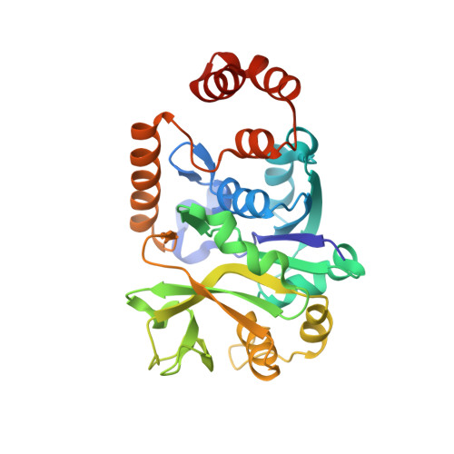
1H5T

Thymidylyltransferase complexed with Thymidylyldiphosphate-glucose


Experimental Data Snapshot

  • Method: X-RAY DIFFRACTION
  • Resolution: 1.90 Å
  • R-Value Free: 0.224 
  • R-Value Work: 0.174 

wwPDB Validation   3D Report Full Report


This is version 2.0 of the entry. See complete history


Literature

Kinetic and Crystallographic Analyses Support a Sequential-Ordered Bi Bi Catalytic Mechanism for Escherichia Coli Glucose-1-Phosphate Thymidylyltransferase

Zuccotti, S.Zanardi, D.Rosano, C.Sturla, L.Tonetti, M.Bolognesi, M.

(2001) J Mol Biol 313: 831

  • DOI: https://doi.org/10.1006/jmbi.2001.5073
  • Primary Citation of Related Structures:  
    1H5R, 1H5S, 1H5T

  • PubMed Abstract: 

    Glucose-1-phosphate thymidylyltransferase is the first enzyme in the biosynthesis of dTDP-l-rhamnose, the precursor of l-rhamnose, an essential component of surface antigens, such as the O-lipopolysaccharide, mediating virulence and adhesion to host tissues in many microorganisms. The enzyme catalyses the formation of dTDP-glucose, from dTTP and glucose 1-phosphate, as well as its pyrophosphorolysis. To shed more light on the catalytic properties of glucose-1-phosphate thymidylyltransferase from Escherichia coli, specifically distinguishing between ping pong and sequential ordered bi bi reaction mechanisms, the enzyme kinetic properties have been analysed in the presence of different substrates and inhibitors. Moreover, three different complexes of glucose-1-phosphate thymidylyltransferase (co-crystallized with dTDP, with dTMP and glucose-1-phosphate, with d-thymidine and glucose-1-phosphate) have been analysed by X-ray crystallography, in the 1.9-2.3 A resolution range (R-factors of 17.3-17.5 %). The homotetrameric enzyme shows strongly conserved substrate/inhibitor binding modes in a surface cavity next to the topological switch-point of a quasi-Rossmann fold. Inspection of the subunit tertiary structure reveals relationships to other enzymes involved in the biosynthesis of nucleotide-sugars, including distant proteins such as the molybdenum cofactor biosynthesis protein MobA. The precise location of the substrate relative to putative reactive residues in the catalytic center suggests that, in keeping with the results of the kinetic measurements, both catalysed reactions, i.e. dTDP-glucose biosynthesis and pyrophosphorolysis, follow a sequential ordered bi bi catalytic mechanism.


  • Organizational Affiliation

    Giannina Gaslini Institute, Largo G. Gaslini, Genova, I-16148, Italy.


Macromolecules
Find similar proteins by:  (by identity cutoff)  |  3D Structure
Entity ID: 1
MoleculeChains Sequence LengthOrganismDetailsImage
Glucose-1-phosphate thymidylyltransferase 1
A, C, D
293Escherichia coliMutation(s): 0 
Gene Names: rfbArmlArmlA1b2039JW2024
EC: 2.7.7.24
UniProt
Find proteins for P37744 (Escherichia coli (strain K12))
Explore P37744 
Go to UniProtKB:  P37744
Entity Groups  
Sequence Clusters30% Identity50% Identity70% Identity90% Identity95% Identity100% Identity
UniProt GroupP37744
Sequence Annotations
Expand
  • Reference Sequence
Find similar proteins by:  (by identity cutoff)  |  3D Structure
Entity ID: 2
MoleculeChains Sequence LengthOrganismDetailsImage
Glucose-1-phosphate thymidylyltransferase 1293Escherichia coliMutation(s): 0 
Gene Names: rfbArmlArmlA1b2039JW2024
EC: 2.7.7.24
UniProt
Find proteins for P37744 (Escherichia coli (strain K12))
Explore P37744 
Go to UniProtKB:  P37744
Entity Groups  
Sequence Clusters30% Identity50% Identity70% Identity90% Identity95% Identity100% Identity
UniProt GroupP37744
Sequence Annotations
Expand
  • Reference Sequence
Small Molecules
Ligands 3 Unique
IDChains Name / Formula / InChI Key2D Diagram3D Interactions
DAU
Query on DAU

Download Ideal Coordinates CCD File 
F [auth A],
H [auth B],
J [auth C],
L [auth D]
2'DEOXY-THYMIDINE-5'-DIPHOSPHO-ALPHA-D-GLUCOSE
C16 H26 N2 O16 P2
YSYKRGRSMLTJNL-URARBOGNSA-N
TYD
Query on TYD

Download Ideal Coordinates CCD File 
E [auth A],
G [auth B],
I [auth C],
K [auth D]
THYMIDINE-5'-DIPHOSPHATE
C10 H16 N2 O11 P2
UJLXYODCHAELLY-XLPZGREQSA-N
SO4
Query on SO4

Download Ideal Coordinates CCD File 
M [auth D]SULFATE ION
O4 S
QAOWNCQODCNURD-UHFFFAOYSA-L
Experimental Data & Validation

Experimental Data

  • Method: X-RAY DIFFRACTION
  • Resolution: 1.90 Å
  • R-Value Free: 0.224 
  • R-Value Work: 0.174 
  • Space Group: P 1 21 1
Unit Cell:
Length ( Å )Angle ( ˚ )
a = 72.81α = 90
b = 119.49β = 112.67
c = 81.17γ = 90
Software Package:
Software NamePurpose
REFMACrefinement
DENZOdata reduction
SCALEPACKdata scaling
EPMRphasing

Structure Validation

View Full Validation Report


Entry History 

Deposition Data

Revision History  (Full details and data files)

  • Version 1.0: 2001-11-23
    Type: Initial release
  • Version 1.1: 2011-05-08
    Changes: Version format compliance
  • Version 1.2: 2011-07-13
    Changes: Version format compliance
  • Version 2.0: 2018-12-05
    Changes: Advisory, Atomic model, Data collection, Database references, Source and taxonomy, Structure summary