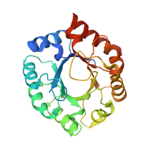
1H1Y

The structure of the cytosolic D-ribulose-5-phosphate 3-epimerase from rice complexed with sulfate


Experimental Data Snapshot

  • Method: X-RAY DIFFRACTION
  • Resolution: 1.87 Å
  • R-Value Free: 0.206 
  • R-Value Work: 0.175 
  • R-Value Observed: 0.175 

wwPDB Validation   3D Report Full Report


This is version 1.4 of the entry. See complete history


Literature

Structure and Catalytic Mechanism of the Cytosolic D-Ribulose-5-Phosphate 3-Epimerase from Rice

Jelakovic, S.Kopriva, S.Suss, K.Schulz, G.E.

(2003) J Mol Biol 326: 127

  • DOI: https://doi.org/10.1016/s0022-2836(02)01374-8
  • Primary Citation of Related Structures:  
    1H1Y, 1H1Z

  • PubMed Abstract: 

    Cytosolic D-ribulose-5-phosphate 3-epimerase from rice was crystallized after EDTA treatment and structurally elucidated by X-ray diffraction to 1.9A resolution. A prominent Zn(2+) site at the active center was established in a soaking experiment. The structure was compared with that of the EDTA-treated crystalline enzyme from the chloroplasts of potato plant leaves showing some structural differences, in particular the "closed" state of a strongly conserved mobile loop covering the substrate at its putative binding site. The previous proposal for the active center was confirmed and the most likely substrate binding position and conformation was derived from the locations of the bound zinc and sulfate ions and of three water molecules. Assuming that the bound zinc ion is an integral part of the enzyme, a reaction mechanism involving a well-stabilized cis-enediolate intermediate is suggested.


  • Organizational Affiliation

    Institut für Organische Chemie und Biochemie, Albert-Ludwigs-Universität, Albertstr. 21, 79104, Freiburg im Breisgau, Germany.


Macromolecules
Find similar proteins by:  (by identity cutoff)  |  3D Structure
Entity ID: 1
MoleculeChains Sequence LengthOrganismDetailsImage
D-RIBULOSE-5-PHOSPHATE 3-EPIMERASE
A, B
228Oryza sativaMutation(s): 0 
EC: 5.1.3.1
UniProt
Find proteins for Q9SE42 (Oryza sativa subsp. japonica)
Explore Q9SE42 
Go to UniProtKB:  Q9SE42
Entity Groups  
Sequence Clusters30% Identity50% Identity70% Identity90% Identity95% Identity100% Identity
UniProt GroupQ9SE42
Sequence Annotations
Expand
  • Reference Sequence
Experimental Data & Validation

Experimental Data

  • Method: X-RAY DIFFRACTION
  • Resolution: 1.87 Å
  • R-Value Free: 0.206 
  • R-Value Work: 0.175 
  • R-Value Observed: 0.175 
  • Space Group: P 1 21 1
Unit Cell:
Length ( Å )Angle ( ˚ )
a = 41.28α = 90
b = 73.3β = 101.75
c = 88.97γ = 90
Software Package:
Software NamePurpose
CNSrefinement

Structure Validation

View Full Validation Report


Entry History 

Deposition Data

Revision History  (Full details and data files)

  • Version 1.0: 2003-01-30
    Type: Initial release
  • Version 1.1: 2011-05-08
    Changes: Version format compliance
  • Version 1.2: 2011-07-13
    Changes: Version format compliance
  • Version 1.3: 2019-07-24
    Changes: Data collection
  • Version 1.4: 2024-05-08
    Changes: Data collection, Database references, Other