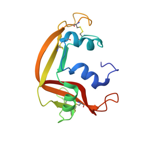
The Crystal Structure of Eosinophil Cationic Protein in Complex with 2'5'-Adp at 2.0 A Resolution Reveals the Details of the Ribonucleolytic Active Site
Mohan, C.G., Boix, E., Evans, H.R., Nikolovski, Z., Nogues, M.V., Cuchillo, C.M., Acharya, K.R.(2002) Biochemistry 41: 12100
- PubMed: 12356310
- DOI: https://doi.org/10.1021/bi0264521
- Primary Citation of Related Structures:
1H1H - PubMed Abstract:
Eosinophil cationic protein (ECP) is a component of the eosinophil granule matrix. It shows marked toxicity against helminth parasites, bacteria single-stranded RNA viruses, and host epithelial cells. Secretion of human ECP is related to eosinophil-associated allergic, asthmatic, and inflammatory diseases. ECP belongs to the pancreatic ribonuclease superfamily of proteins, and the crystal structure of ECP in the unliganded form (determined previously) exhibited a conserved RNase A fold [Boix, E., et al. (1999) Biochemistry 38, 16794-16801]. We have now determined a high-resolution (2.0 A) crystal structure of ECP in complex with adenosine 2',5'-diphosphate (2',5'-ADP) which has revealed the details of the ribonucleolytic active site. Residues Gln-14, His-15, and Lys-38 make hydrogen bond interactions with the phosphate at the P(1) site, while His-128 interacts with the purine ring at the B(2) site. A new phosphate binding site, P(-)(1), has been identified which involves Arg-34. This study is the first detailed structural analysis of the nucleotide recognition site in ECP and provides a starting point for the understanding of its substrate specificity and low catalytic efficiency compared with that of the eosinophil-derived neurotoxin (EDN), a close homologue.
- Department of Biology and Biochemistry, University of Bath, Claverton Down, Bath BA2 7AY, U.K.
Organizational Affiliation:


