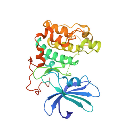
1GZO

Structure of protein kinase B unphosphorylated


Experimental Data Snapshot

  • Method: X-RAY DIFFRACTION
  • Resolution: 2.75 Å
  • R-Value Free: 0.310 
  • R-Value Work: 0.248 
  • R-Value Observed: 0.248 

Starting Model: experimental
View more details

wwPDB Validation   3D Report Full Report


This is version 1.3 of the entry. See complete history


Literature

Molecular Mechanism for the Regulation of Protein Kinase B/Akt by Hydrophobic Motif Phosphorylation

Yang, J.Cron, P.Thompson, V.Good, V.Hess, D.Hemmings, B.A.Barford, D.

(2002) Mol Cell 9: 1227

  • DOI: https://doi.org/10.1016/s1097-2765(02)00550-6
  • Primary Citation of Related Structures:  
    1GZK, 1GZN, 1GZO

  • PubMed Abstract: 

    Protein kinase B/Akt plays crucial roles in promoting cell survival and mediating insulin responses. The enzyme is stimulated by phosphorylation at two regulatory sites: Thr 309 of the activation segment and Ser 474 of the hydrophobic motif, a conserved feature of many AGC kinases. Analysis of the crystal structures of the unphosphorylated and Thr 309 phosphorylated states of the PKB kinase domain provides a molecular explanation for regulation by Ser 474 phosphorylation. Activation by Ser 474 phosphorylation occurs via a disorder to order transition of the alphaC helix with concomitant restructuring of the activation segment and reconfiguration of the kinase bilobal structure. These conformational changes are mediated by a phosphorylation-promoted interaction of the hydrophobic motif with a channel on the N-terminal lobe induced by the ordered alphaC helix and are mimicked by peptides corresponding to the hydrophobic motif of PKB and potently by the hydrophobic motif of PRK2.


  • Organizational Affiliation

    Section of Structural Biology, Institute of Cancer Research, Chester Beatty Laboratories, 237 Fulham Road, London SW3 6JB, UK.


Macromolecules
Find similar proteins by:  (by identity cutoff)  |  3D Structure
Entity ID: 1
MoleculeChains Sequence LengthOrganismDetailsImage
RAC-BETA SERINE/THREONINE PROTEIN KINASE315Homo sapiensMutation(s): 0 
EC: 2.7.1 (PDB Primary Data), 2.7.11.1 (UniProt)
UniProt & NIH Common Fund Data Resources
Find proteins for P31751 (Homo sapiens)
Explore P31751 
Go to UniProtKB:  P31751
PHAROS:  P31751
GTEx:  ENSG00000105221 
Entity Groups  
Sequence Clusters30% Identity50% Identity70% Identity90% Identity95% Identity100% Identity
UniProt GroupP31751
Sequence Annotations
Expand
  • Reference Sequence
Experimental Data & Validation

Experimental Data

  • Method: X-RAY DIFFRACTION
  • Resolution: 2.75 Å
  • R-Value Free: 0.310 
  • R-Value Work: 0.248 
  • R-Value Observed: 0.248 
  • Space Group: P 41 21 2
Unit Cell:
Length ( Å )Angle ( ˚ )
a = 149.703α = 90
b = 149.703β = 90
c = 39.185γ = 90
Software Package:
Software NamePurpose
CNSrefinement
DENZOdata reduction
SCALEPACKdata scaling
CNSphasing

Structure Validation

View Full Validation Report


Entry History 

Deposition Data

Revision History  (Full details and data files)

  • Version 1.0: 2003-05-22
    Type: Initial release
  • Version 1.1: 2011-05-08
    Changes: Version format compliance
  • Version 1.2: 2011-07-13
    Changes: Version format compliance
  • Version 1.3: 2023-12-13
    Changes: Data collection, Database references, Other, Refinement description