1GUJ

Insulin at pH 2: structural analysis of the conditions promoting insulin fibre formation.


Experimental Data Snapshot

  • Method: X-RAY DIFFRACTION
  • Resolution: 1.62 Å
  • R-Value Free: 0.207 
  • R-Value Work: 0.170 
  • R-Value Observed: 0.172 

wwPDB Validation   3D Report Full Report


This is version 1.4 of the entry. See complete history


Literature

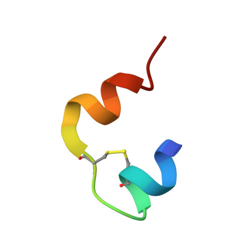
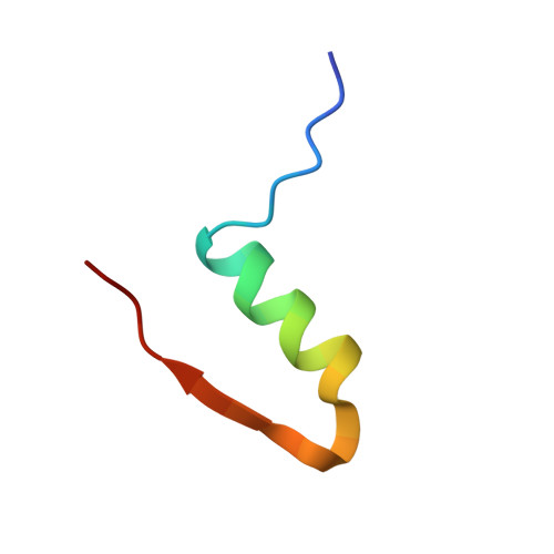
Insulin at Ph2: Structural Analysis of the Conditions Promoting Insulin Fibre Formation

Whittingham, J.L.Scott, D.J.Chance, K.Wilson, A.Finch, J.Brange, J.Dodson, G.G.

(2002) J Mol Biol 318: 479

  • DOI: https://doi.org/10.1016/S0022-2836(02)00021-9
  • Primary Citation of Related Structures:  
    1GUJ

  • PubMed Abstract: 

    When insulin solutions are subjected to acid, heat and agitation, the normal pattern of insulin assembly (dimers-->tetramers-->hexamers) is disrupted; the molecule undergoes conformational changes allowing it to follow an alternative aggregation pathway (via a monomeric species) leading to the formation of insoluble amyloid fibres. To investigate the effect of acid pH on the conformation and aggregation state of the protein, the crystal structure of human insulin at pH 2.1 has been determined to 1.6 A resolution. The structure reveals that the native fold is maintained at low pH, and that the molecule is still capable of forming dimers similar to those found in hexameric insulin structures at higher pH. Sulphate ions are incorporated into the molecule and the crystal lattice where they neutralise positive charges on the protein, stabilising its structure and facilitating crystallisation. The sulphate interactions are associated with local deformations in the protein, which may indicate that the structure is more plastic at low pH. Transmission electron microscopy analysis of insulin fibres reveals that the appearance of the fibres is greatly influenced by the type of acid employed. Sulphuric acid produces distinctive highly bunched, truncated fibres, suggesting that the sulphate ions have a sophisticated role to play in fibre formation, rather as they do in the crystal structure. Analytical ultracentrifugation studies show that in the absence of heating, insulin is predominantly dimeric in mineral acids, whereas in acetic acid the equilibrium is shifted towards the monomer. Hence, the effect of acid on the aggregation state of insulin is also complex. These results suggest that acid conditions increase the susceptibility of the molecule to conformational change and dissociation, and enhance the rate of fibrillation by providing a charged environment in which the attractive forces between the protein molecules is increased.


  • Organizational Affiliation

    York Structural Biology Laboratory, Department of Chemistry, University of York, Heslington, York YO10 5DD, UK. jean@ysbl.york.ac.uk


Macromolecules

Find similar proteins by:  Sequence   |   3D Structure  

Entity ID: 1
MoleculeChains Sequence LengthOrganismDetailsImage
INSULIN
A, C
21Homo sapiensMutation(s): 0 
UniProt & NIH Common Fund Data Resources
Find proteins for P01308 (Homo sapiens)
Explore P01308 
Go to UniProtKB:  P01308
PHAROS:  P01308
GTEx:  ENSG00000254647 
Entity Groups  
Sequence Clusters30% Identity50% Identity70% Identity90% Identity95% Identity100% Identity
UniProt GroupP01308
Sequence Annotations
Expand
  • Reference Sequence
Find similar proteins by:  (by identity cutoff)  |  3D Structure
Entity ID: 2
MoleculeChains Sequence LengthOrganismDetailsImage
INSULIN
B, D
30Homo sapiensMutation(s): 0 
UniProt & NIH Common Fund Data Resources
Find proteins for P01308 (Homo sapiens)
Explore P01308 
Go to UniProtKB:  P01308
PHAROS:  P01308
GTEx:  ENSG00000254647 
Entity Groups  
Sequence Clusters30% Identity50% Identity70% Identity90% Identity95% Identity100% Identity
UniProt GroupP01308
Sequence Annotations
Expand
  • Reference Sequence
Small Molecules
Ligands 1 Unique
IDChains Name / Formula / InChI Key2D Diagram3D Interactions
SO4
Query on SO4

Download Ideal Coordinates CCD File 
E [auth A],
F [auth D]
SULFATE ION
O4 S
QAOWNCQODCNURD-UHFFFAOYSA-L
Experimental Data & Validation

Experimental Data

  • Method: X-RAY DIFFRACTION
  • Resolution: 1.62 Å
  • R-Value Free: 0.207 
  • R-Value Work: 0.170 
  • R-Value Observed: 0.172 
  • Space Group: P 21 21 21
Unit Cell:
Length ( Å )Angle ( ˚ )
a = 44.046α = 90
b = 46.177β = 90
c = 51.194γ = 90
Software Package:
Software NamePurpose
REFMACrefinement
DENZOdata reduction
SCALEPACKdata scaling
AMoREphasing

Structure Validation

View Full Validation Report


Entry History 

Deposition Data

Revision History  (Full details and data files)

  • Version 1.0: 2002-03-08
    Type: Initial release
  • Version 1.1: 2011-05-08
    Changes: Version format compliance
  • Version 1.2: 2011-07-13
    Changes: Version format compliance
  • Version 1.3: 2019-05-08
    Changes: Advisory, Data collection, Experimental preparation, Other
  • Version 1.4: 2023-12-13
    Changes: Advisory, Data collection, Database references, Refinement description