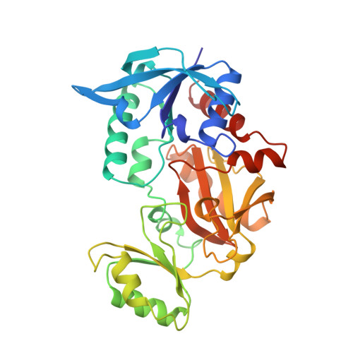
A pseudo-michaelis quaternary complex in the reverse reaction of a ligase: structure of Escherichia coli B glutathione synthetase complexed with ADP, glutathione, and sulfate at 2.0 A resolution.
Hara, T., Kato, H., Katsube, Y., Oda, J.(1996) Biochemistry 35: 11967-11974
- PubMed: 8810901
- DOI: https://doi.org/10.1021/bi9605245
- Primary Citation of Related Structures:
1GSA - PubMed Abstract:
The crystal structure of glutathione synthetase from Escherichia coli B complexed with ADP, glutathione, and sulfate has been determined at 2.0 A resolution. Concerning the chemical similarity of sulfate and phosphate, this quaternary complex structure represents a pseudo enzyme-substrate complex in the reverse reaction and consequently allows us to understand the active site architecture of the E. coli glutathione synthetase. Two Mg2+ ions are coordinated with oxygen atoms from the alpha- and beta-phosphate groups of ADP and from the sulfate ion. The flexible loops, invisible in the unliganded or the binary and ternary complex structures, are fixed in the quaternary complex. The larger flexible loop (Ile226-Arg241) includes one turn of a 310-helix that comprises the binding site of the glycine moiety of GSH. The small loop (Gly164-Gly167) is involved in nucleotide binding and acts as a phosphate gripper. The side chains of Arg210 and Arg225 interact with the sulfate ion and the beta-phosphate moiety of ADP. Arg 210 is likely to interact with the carboxylate of the C-terminal gamma-glutamylcysteine in the substrate-binding form of the forward reaction. Other positively charged residues in the active site (Lys125 and Lys160) are involved in nucleotide binding, directing the phosphate groups to the right position for catalysis. Functional aspects of the active site architecture in the substrate-binding form are discussed.
- Institute for Chemical Research, Kyoto University, Japan.
Organizational Affiliation:


