1GS4

Structural basis for the glucocorticoid response in a mutant human androgen receptor (ARccr) derived from an androgen-independent prostate cancer


Experimental Data Snapshot

  • Method: X-RAY DIFFRACTION
  • Resolution: 1.95 Å
  • R-Value Free: 0.285 
  • R-Value Observed: 0.205 

wwPDB Validation   3D Report Full Report


Ligand Structure Quality Assessment 


This is version 2.1 of the entry. See complete history


Literature

Structural Basis for the Glucocorticoid Response in a Mutant Human Androgen Receptor (Ar(Ccr)) Derived from an Androgen-Independent Prostate Cancer

Matias, P.M.Carrondo, M.A.Coelho, R.Thomaz, M.Zhao, X.-Y.Wegg, A.Crusius, K.Egner, U.Donner, P.

(2002) J Med Chem 45: 1439-1446

  • DOI: https://doi.org/10.1021/jm011072j
  • Primary Citation of Related Structures:  
    1GS4

  • PubMed Abstract: 

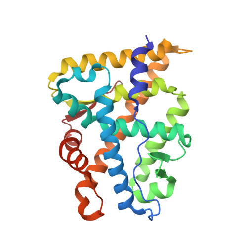
    The crystal structure of a mutant androgen receptor (AR) ligand-binding domain (LBD) in complex with the agonist 9alpha-fluorocortisol has been determined at 1.95 A resolution. This mutant AR contains two mutations (L701H and T877A) and was previously reported as a high-affinity cortisol/cortisone responsive AR (AR(ccr)) isolated from the androgen-independent human prostate cancer cell lines MDA PCa 2a and 2b (Zhao et al. Nature Med. 2000, 6, 703-6). The three-dimensional structure of the AR(ccr) LBD complexed with 9alpha-fluorocortisol shows the typical conformation of an agonist-bound nuclear receptor in which helix 12 is precisely positioned as a "lid" for the ligand-binding pocket. Binding of 9alpha-fluorocortisol to the AR(ccr) involves favorable hydrogen bond patterns on the C17 and C21 substituents of the ligand due to the mutations at 701 and 877 in the AR(ccr). Our studies provide the first structural explanation for the glucocorticoid activation of AR(ccr), which is important for the development of new therapeutic treatments for androgen-independent prostate cancer.


  • Organizational Affiliation

    Instituto de Tecnologia Química e Biológica, Universidade Nova de Lisboa, Apartado 127, 2780 Oeiras, Portugal.


Macromolecules
Find similar proteins by:  (by identity cutoff)  |  3D Structure
Entity ID: 1
MoleculeChains Sequence LengthOrganismDetailsImage
ANDROGEN RECEPTOR248Homo sapiensMutation(s): 2 
UniProt & NIH Common Fund Data Resources
Find proteins for P10275 (Homo sapiens)
Explore P10275 
Go to UniProtKB:  P10275
PHAROS:  P10275
GTEx:  ENSG00000169083 
Entity Groups  
Sequence Clusters30% Identity50% Identity70% Identity90% Identity95% Identity100% Identity
UniProt GroupP10275
Sequence Annotations
Expand
  • Reference Sequence
Small Molecules
Ligands 2 Unique
IDChains Name / Formula / InChI Key2D Diagram3D Interactions
ZK5
Query on ZK5

Download Ideal Coordinates CCD File 
B [auth A]9ALPHA-FLUOROCORTISOL
C21 H29 F O5
AAXVEMMRQDVLJB-BULBTXNYSA-N
PO4
Query on PO4

Download Ideal Coordinates CCD File 
C [auth A]PHOSPHATE ION
O4 P
NBIIXXVUZAFLBC-UHFFFAOYSA-K
Experimental Data & Validation

Experimental Data

  • Method: X-RAY DIFFRACTION
  • Resolution: 1.95 Å
  • R-Value Free: 0.285 
  • R-Value Observed: 0.205 
  • Space Group: P 21 21 21
Unit Cell:
Length ( Å )Angle ( ˚ )
a = 56.35α = 90
b = 66.23β = 90
c = 73.05γ = 90
Software Package:
Software NamePurpose
SHELXL-97refinement
DENZOdata reduction
SCALEPACKdata scaling
AMoREphasing

Structure Validation

View Full Validation Report


Ligand Structure Quality Assessment 


Entry History 

Deposition Data

Revision History  (Full details and data files)

  • Version 1.0: 2003-01-16
    Type: Initial release
  • Version 1.1: 2011-05-08
    Changes: Version format compliance
  • Version 1.2: 2011-07-13
    Changes: Version format compliance
  • Version 2.0: 2018-02-28
    Changes: Advisory, Atomic model, Database references, Source and taxonomy
  • Version 2.1: 2023-12-13
    Changes: Advisory, Data collection, Database references, Other, Refinement description