Crystallographic binding studies on the allosteric inhibitor glucose-6-phosphate to T state glycogen phosphorylase b.
Johnson, L.N., Snape, P., Martin, J.L., Acharya, K.R., Barford, D., Oikonomakos, N.G.(1993) J Mol Biology 232: 253-267
- PubMed: 8331662 Search on PubMed
- DOI: https://doi.org/10.1006/jmbi.1993.1380
- Primary Citation Related Structures:
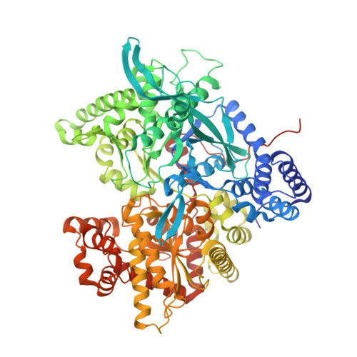
1GPY - PubMed Abstract:
Glucose-6-phosphate is an important allosteric inhibitor of glycogen phosphorylase b that restrains the enzyme in the inactive state in resting muscle. A crystallographic binding study by diffusion of glucose-6-phosphate into performed crystals of T state phosphorylase b has been carried out at 2.3 A resolution and the structure refined by restrained crystallographic least-squares and simulated annealing to give a crystallographic R-value of 0.203. The inhibitor binds at the AMP allosteric effector site at the subunit-subunit interface of the dimer. The phosphate groups of the glucose-6-phosphate and AMP occupy partially overlapping sites and make similar contacts to two arginine residues (Arg309 and Arg310) but in glucose-6-phosphate there is a contact to a third arginine (Arg242). The glucopyranose of glucose-6-phosphate and the adenine ribose of AMP occupy different positions. Including the contacts to the three arginine residues by the phosphate group, the glucose-6-phosphate makes a total of 11 hydrogen-bonds to the enzyme and all but one of these are to charged groups. The O-2 hydroxyl hydrogen-bonds to the main-chain carbonyl oxygen of Val40' from the other subunit and this interaction appears important for the allosteric response. There are substantial conformational changes both in the vicinity of the glucose-6-phosphate (involving for example Phe196 and Arg309) and at the subunit interface (involving residues 42' to 51' and 192 to 196). These shifts tighten the binding of the inhibitor and the interface. Comparison of the glucose-6-phosphate complex with the T state native phosphorylase b and the R state phosphorylase a structures shows that there is a graded response from T state glucose-6-phosphate complex through T state phosphorylase b to R state phosphorylase a that suggests that glucose-6-phosphate promotes a tight structure that is more "tensed" than native T state phosphorylase b. The results show how the same allosteric effector site can exhibit a tight binding site for the activator AMP in the R state structure and a tight binding site for glucose-6-phosphate in the modified T state structure.
- Laboratory of Molecular Biophysics, University of Oxford, U.K.
Organizational Affiliation: