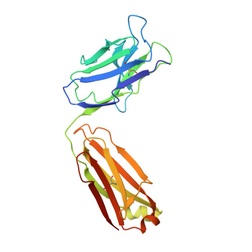
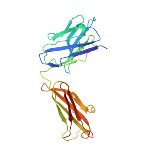
1GGI

CRYSTAL STRUCTURE OF AN HIV-1 NEUTRALIZING ANTIBODY 50.1 IN COMPLEX WITH ITS V3 LOOP PEPTIDE ANTIGEN


Experimental Data Snapshot

  • Method: X-RAY DIFFRACTION
  • Resolution: 2.80 Å
  • R-Value Work: 0.188 
  • R-Value Observed: 0.188 

wwPDB Validation   3D Report Full Report


This is version 1.4 of the entry. See complete history


Literature

Crystal structure of a human immunodeficiency virus type 1 neutralizing antibody, 50.1, in complex with its V3 loop peptide antigen.

Rini, J.M.Stanfield, R.L.Stura, E.A.Salinas, P.A.Profy, A.T.Wilson, I.A.

(1993) Proc Natl Acad Sci U S A 90: 6325-6329

  • DOI: https://doi.org/10.1073/pnas.90.13.6325
  • Primary Citation of Related Structures:  
    1GGI

  • PubMed Abstract: 

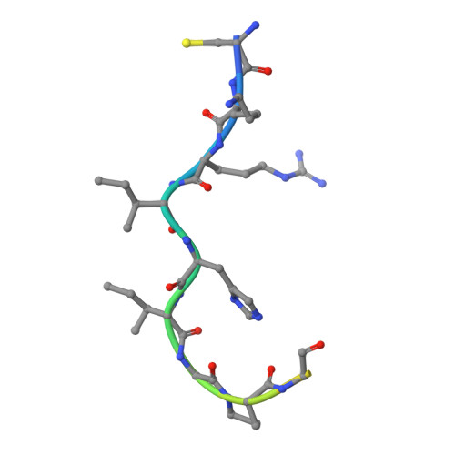
    The crystal structure of the Fab fragment of a human immunodeficiency virus type 1 (HIV-1) neutralizing monoclonal antibody Fab has been determined at 2.8 A resolution in complex with a linear 16-residue peptide from the third hypervariable region (V3) of gp120. The first 9 residues of the peptide are ordered in the electron density maps, and their conformation is in partial agreement with the beta-strand-type II beta-turn structure predicted for this portion of the V3 loop. Notably, several of the peptide residues that are well conserved among different HIV-1 isolates contact a nonpolar 25-A-long groove in the antibody-combining site. The largely extended structure of the peptide differs from the beta-turns seen as the primary determinants in other published anti-peptide Fab structures. Analysis of the specific Fab-peptide interactions only partially explains the MN isolate specificity shown by this antibody.


  • Organizational Affiliation

    Department of Molecular Biology, Scripps Research Institute, La Jolla, CA 92037.


Macromolecules
Find similar proteins by:  (by identity cutoff)  |  3D Structure
Entity ID: 1
MoleculeChains Sequence LengthOrganismDetailsImage
IGG2A 50.1 FAB (LIGHT CHAIN)A [auth L],
D [auth M]
218Mus musculusMutation(s): 0 
Entity Groups  
Sequence Clusters30% Identity50% Identity70% Identity90% Identity95% Identity100% Identity
Sequence Annotations
Expand
  • Reference Sequence
Find similar proteins by:  (by identity cutoff)  |  3D Structure
Entity ID: 2
MoleculeChains Sequence LengthOrganismDetailsImage
IGG2A 50.1 FAB (HEAVY CHAIN)B [auth H],
E [auth J]
222Mus musculusMutation(s): 0 
Gene Names: Ighg
UniProt
Find proteins for P01863 (Mus musculus)
Explore P01863 
Go to UniProtKB:  P01863
Entity Groups  
Sequence Clusters30% Identity50% Identity70% Identity90% Identity95% Identity100% Identity
UniProt GroupP01863
Sequence Annotations
Expand
  • Reference Sequence

Find similar proteins by:  Sequence   |   3D Structure  

Entity ID: 3
MoleculeChains Sequence LengthOrganismDetailsImage
HIV-1 V3 LOOP PEPTIDE ANTIGENC [auth P],
F [auth Q]
16Human immunodeficiency virus type 1 group M subtype B (isolate MN)Mutation(s): 0 
Gene Names: env
UniProt
Find proteins for P05877 (Human immunodeficiency virus type 1 group M subtype B (isolate MN))
Explore P05877 
Go to UniProtKB:  P05877
Entity Groups  
Sequence Clusters30% Identity50% Identity70% Identity90% Identity95% Identity100% Identity
UniProt GroupP05877
Sequence Annotations
Expand
  • Reference Sequence
Experimental Data & Validation

Experimental Data

  • Method: X-RAY DIFFRACTION
  • Resolution: 2.80 Å
  • R-Value Work: 0.188 
  • R-Value Observed: 0.188 
  • Space Group: P 1 21 1
Unit Cell:
Length ( Å )Angle ( ˚ )
a = 130.33α = 90
b = 52.57β = 97.5
c = 82.04γ = 90
Software Package:
Software NamePurpose
X-PLORmodel building
X-PLORrefinement
X-PLORphasing

Structure Validation

View Full Validation Report


Entry History 

Deposition Data

Revision History  (Full details and data files)

  • Version 1.0: 1993-10-31
    Type: Initial release
  • Version 1.1: 2008-03-24
    Changes: Version format compliance
  • Version 1.2: 2011-07-13
    Changes: Version format compliance
  • Version 1.3: 2024-06-05
    Changes: Data collection, Database references, Derived calculations, Other
  • Version 1.4: 2024-10-16
    Changes: Structure summary