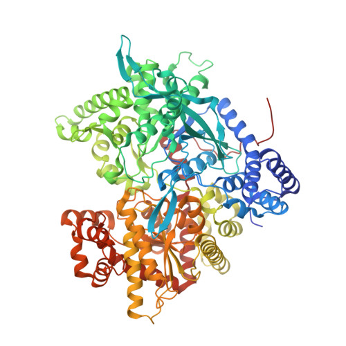
Design of inhibitors of glycogen phosphorylase: a study of alpha- and beta-C-glucosides and 1-thio-beta-D-glucose compounds.
Watson, K.A., Mitchell, E.P., Johnson, L.N., Son, J.C., Bichard, C.J., Orchard, M.G., Fleet, G.W., Oikonomakos, N.G., Leonidas, D.D., Kontou, M., Papageorgioui, A.(1994) Biochemistry 33: 5745-5758
- PubMed: 8180201
- DOI: https://doi.org/10.1021/bi00185a011
- Primary Citation of Related Structures:
1GG8 - PubMed Abstract:
alpha-D-Glucose is a weak inhibitor of glycogen phosphorylase b (Ki = 1.7 mM) and acts as a physiological regulator of hepatic glycogen metabolism. Glucose binds to phosphorylase at the catalytic site and results in a conformational change that stabilizes the inactive T state of the enzyme, promoting the action of protein phosphatase 1 and stimulating glycogen synthase. It has been suggested that, in the liver, glucose analogues with greater affinity for glycogen phosphorylase may result in a more effective regulatory agent. Several alpha- and beta-anhydroglucoheptonic acid derivatives and 1-deoxy-1-thio-beta-D-glucose analogues have been synthesized and tested in a series of crystallographic and kinetic binding studies with glycogen phosphorylase. The structural results of the bound enzyme-ligand complexes have been analyzed, together with the resulting affinities, in an effort to understand and exploit the molecular interactions that might give rise to a better inhibitor. This work has shown the following: (i) Similar affinities may be obtained through different sets of interactions. Specifically, in the case of the alpha- and beta-glucose-C-amides, similar Ki's (0.37 and 0.44 mM, respectively) are obtained with the alpha-anomer through interactions from the ligand via water molecules to the protein and with the beta-anomer through direct interaction from the ligand to the protein. Thus, hydrogen bonds through water can contribute binding energy similar to that of hydrogen bonds directly to the protein. (ii) Attempts to improve the inhibition by additional groups did not always lead to the expected result. The addition of nonpolar groups to the alpha-carboxamide resulted in a change in conformation of the pyranose ring from a chair to a skew boat and the consequent loss of favorable hydrogen bonds and increase in the Ki. (iii) The addition of polar groups to the alpha-carboxamide led to compounds with the chair conformation, and in the examples studied, it appears that hydration by a water molecule may provide sufficient stabilization to retain the chair conformation. (iv) The best inhibitor was N-methyl-beta-glucose-C-carboxamide (Ki = 0.16 mM), which showed a 46-fold improvement in Ki from the parent beta-D-glucose. The decrease in Ki may be accounted for by a single hydrogen bond from the amide nitrogen to a main-chain carbonyl oxygen, an increase in entropy through displacement of a water molecule, and favorable van der Waals contacts between the methyl substituent and nonpolar protein residues.(ABSTRACT TRUNCATED AT 250 WORDS)
- Oxford Centre for Molecular Sciences, U.K.
Organizational Affiliation:


