1G8Y | pdb_00001g8y

CRYSTAL STRUCTURE OF THE HEXAMERIC REPLICATIVE HELICASE REPA OF PLASMID RSF1010


Experimental Data Snapshot

  • Method: X-RAY DIFFRACTION
  • Resolution: 2.40 Å
  • R-Value Free: 
    0.264 (Depositor) 
  • R-Value Work: 
    0.221 (Depositor) 

wwPDB Validation 3D Report Full Report

Validation slider image for 1G8Y

This is version 1.3 of the entry. See complete history

Literature

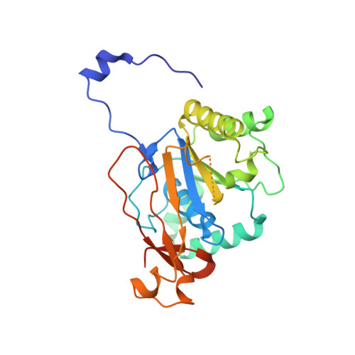
Crystal structure of the hexameric replicative helicase RepA of plasmid RSF1010.

Niedenzu, T.Roleke, D.Bains, G.Scherzinger, E.Saenger, W.

(2001) J Mol Biology 306: 479-487

  • DOI: https://doi.org/10.1006/jmbi.2000.4398
  • Primary Citation Related Structures: 
    1G8Y

  • PubMed Abstract: 

    Unwinding of double-stranded DNA into single-stranded intermediates required for various fundamental life processes is catalyzed by helicases, a family of mono-, di- or hexameric motor proteins fueled by nucleoside triphosphate hydrolysis. The three-dimensional crystal structure of the hexameric helicase RepA encoded by plasmid RSF1010 has been determined by X-ray diffraction at 2.4 A resolution. The hexamer shows an annular structure with 6-fold rotational symmetry and a approximately 17 A wide central hole, suggesting that single-stranded DNA may be threaded during unwinding. Homologs of all five conserved sequence motifs of the DnaB-like helicase family are found in RepA, and the topography of the monomer resembles RecA and the helicase domain of the bacteriophage T7 gp4 protein. In a modeled complex, ATP molecules are located at the subunit interfaces and clearly define adenine-binding and ATPase catalytic sites formed by amino acid residues located on adjacent monomers; most remarkable is the "arginine finger" Arg207 contributing to the active site in the adjacent monomer. This arrangement of active-site residues suggests cooperativity between monomers in ATP hydrolysis and helicase activity of RepA. The mechanism of DNA unwinding remains elusive, as RepA is 6-fold symmetric, contrasting the recently published asymmetric structure of the bacteriophage T7 gp4 helicase domain.


  • Organizational Affiliation
    • Institut für Kristallographie, Freie Universität Berlin, Takustr. 6, Berlin, D-14195, Germany.

Macromolecule Content 

  • Total Structure Weight: 359.34 kDa 
  • Atom Count: 22,536 
  • Modeled Residue Count: 2,904 
  • Deposited Residue Count: 3,348 
  • Unique protein chains: 1

Macromolecules

Find similar proteins by:|  3D Structure
Entity ID: 1
MoleculeChains  Sequence LengthOrganismDetailsImage
REGULATORY PROTEIN REPA
A, B, C, D, E
A, B, C, D, E, F, G, H, I, J, K, L
279Escherichia coliMutation(s): 0 
Gene Names: REPA ON PLASMID INCQ RSF1010
UniProt
Find proteins for P20356 (Escherichia coli)
Explore P20356 
Go to UniProtKB:  P20356
Entity Groups
Sequence Clusters30% Identity50% Identity70% Identity90% Identity95% Identity100% Identity
UniProt GroupP20356
Sequence Annotations
Expand
Reference Sequence

Experimental Data & Validation

Experimental Data

  • Method: X-RAY DIFFRACTION
  • Resolution: 2.40 Å
  • R-Value Free:  0.264 (Depositor) 
  • R-Value Work:  0.221 (Depositor) 
Space Group: P 1 21 1
Unit Cell:
Length ( Å )Angle ( ˚ )
a = 104.432α = 90
b = 179.156β = 108.8
c = 116.289γ = 90
Software Package:
Software NamePurpose
MLPHAREphasing
CNSrefinement
DENZOdata reduction
SCALEPACKdata scaling

Structure Validation

View Full Validation Report


Entry History 

Deposition Data

Revision History  (Full details and data files)

  • Version 1.0: 2001-02-14
    Type: Initial release
  • Version 1.1: 2008-04-27
    Changes: Version format compliance
  • Version 1.2: 2011-07-13
    Changes: Version format compliance
  • Version 1.3: 2024-02-07
    Changes: Data collection, Database references