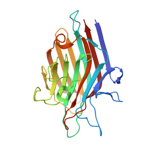
1G8W

IMPROVED STRUCTURE OF PHYTOHEMAGGLUTININ-L FROM THE KIDNEY BEAN


Experimental Data Snapshot

  • Method: X-RAY DIFFRACTION
  • Resolution: 2.80 Å
  • R-Value Free: 0.212 
  • R-Value Work: 0.181 
  • R-Value Observed: 0.181 

Starting Model: experimental
View more details

wwPDB Validation   3D Report Full Report


Ligand Structure Quality Assessment 


This is version 1.7 of the entry. See complete history


Literature

Weak protein-protein interactions in lectins: the crystal structure of a vegetative lectin from the legume Dolichos biflorus.

Buts, L.Dao-Thi, M.H.Loris, R.Wyns, L.Etzler, M.Hamelryck, T.

(2001) J Mol Biol 309: 193-201

  • DOI: https://doi.org/10.1006/jmbi.2001.4639
  • Primary Citation of Related Structures:  
    1G7Y, 1G8W, 1G9F

  • PubMed Abstract: 

    The legume lectins are widely used as a model system for studying protein-carbohydrate and protein-protein interactions. They exhibit a fascinating quaternary structure variation, which becomes important when they interact with multivalent glycoconjugates, for instance those on cell surfaces. Recently, it has become clear that certain lectins form weakly associated oligomers. This phenomenon may play a role in the regulation of receptor crosslinking and subsequent signal transduction. The crystal structure of DB58, a dimeric lectin from the legume Dolichos biflorus reveals a separate dimer of a previously unobserved type, in addition to a tetramer consisting of two such dimers. This tetramer resembles that formed by DBL, the seed lectin from the same plant. A single amino acid substitution in DB58 affects the conformation and flexibility of a loop in the canonical dimer interface. This disrupts the formation of a stable DBL-like tetramer in solution, but does not prohibit its formation in suitable conditions, which greatly increases the possibilities for the cross-linking of multivalent ligands. The non-canonical DB58 dimer has a buried symmetrical alpha helix, which can be present in the crystal in either of two antiparallel orientations. Two existing structures and datasets for lectins with similar quaternary structures were reconsidered. A central alpha helix could be observed in the soybean lectin, but not in the leucoagglutinating lectin from Phaseolus vulgaris. The relative position and orientation of the carbohydrate-binding sites in the DB58 dimer may affect its ability to crosslink mulitivalent ligands, compared to the other legume lectin dimers.


  • Organizational Affiliation

    ULTR-Ultrastructure Department, Vrije Universiteit Brussel, Sint-Genesius-Rode Belgium. lieven@ultr.vub.ac.be


Macromolecules
Find similar proteins by:  (by identity cutoff)  |  3D Structure
Entity ID: 1
MoleculeChains Sequence LengthOrganismDetailsImage
LEUCOAGGLUTINATING PHYTOHEMAGGLUTININ
A, B, C, D
233Phaseolus vulgarisMutation(s): 0 
UniProt
Find proteins for P05087 (Phaseolus vulgaris)
Explore P05087 
Go to UniProtKB:  P05087
Entity Groups  
Sequence Clusters30% Identity50% Identity70% Identity90% Identity95% Identity100% Identity
UniProt GroupP05087
Glycosylation
Glycosylation Sites: 1
Sequence Annotations
Expand
  • Reference Sequence
Small Molecules
Ligands 3 Unique
IDChains Name / Formula / InChI Key2D Diagram3D Interactions
NAG
Query on NAG

Download Ideal Coordinates CCD File 
E [auth A],
H [auth B],
K [auth C],
N [auth D]
2-acetamido-2-deoxy-beta-D-glucopyranose
C8 H15 N O6
OVRNDRQMDRJTHS-FMDGEEDCSA-N
MN
Query on MN

Download Ideal Coordinates CCD File 
F [auth A],
I [auth B],
L [auth C],
O [auth D]
MANGANESE (II) ION
Mn
WAEMQWOKJMHJLA-UHFFFAOYSA-N
CA
Query on CA

Download Ideal Coordinates CCD File 
G [auth A],
J [auth B],
M [auth C],
P [auth D]
CALCIUM ION
Ca
BHPQYMZQTOCNFJ-UHFFFAOYSA-N
Experimental Data & Validation

Experimental Data

  • Method: X-RAY DIFFRACTION
  • Resolution: 2.80 Å
  • R-Value Free: 0.212 
  • R-Value Work: 0.181 
  • R-Value Observed: 0.181 
  • Space Group: C 1 2 1
Unit Cell:
Length ( Å )Angle ( ˚ )
a = 106.479α = 90
b = 121.278β = 93.18
c = 89.957γ = 90
Software Package:
Software NamePurpose
AMoREphasing
CNSrefinement
MADNESSdata reduction

Structure Validation

View Full Validation Report


Ligand Structure Quality Assessment 


Entry History 

Deposition Data

Revision History  (Full details and data files)

  • Version 1.0: 2000-12-13
    Type: Initial release
  • Version 1.1: 2008-04-27
    Changes: Version format compliance
  • Version 1.2: 2011-07-13
    Changes: Non-polymer description, Version format compliance
  • Version 1.3: 2017-10-04
    Changes: Refinement description
  • Version 1.4: 2018-04-18
    Changes: Data collection
  • Version 1.5: 2020-07-29
    Type: Remediation
    Reason: Carbohydrate remediation
    Changes: Data collection, Derived calculations, Structure summary
  • Version 1.6: 2023-08-09
    Changes: Data collection, Database references, Derived calculations, Refinement description, Structure summary
  • Version 1.7: 2024-10-16
    Changes: Structure summary