1G8N | pdb_00001g8n

MOLECULAR AND CRYSTAL STRUCTURE OF D(CGCGAATF5UCGCG):5-FORMYLURIDINE/ ADENOSINE BASE-PAIRS IN B-DNA


Experimental Data Snapshot

  • Method: X-RAY DIFFRACTION
  • Resolution: 1.55 Å
  • R-Value Free: 
    0.232 (Depositor) 
  • R-Value Work: 
    0.207 (Depositor) 

Starting Model: experimental
View more details

wwPDB Validation 3D Report Full Report

Validation slider image for 1G8N

This is version 1.3 of the entry. See complete history

Literature

Crystallization and preliminary X-ray analysis of a DNA dodecamer containing 2'-deoxy-5-formyluridine; what is the role of magnesium cation in crystallization of Dickerson-type DNA dodecamers?

Tsunoda, M.Karino, N.Ueno, Y.Matsuda, A.Takenaka, A.

(2001) Acta Crystallogr D Biol Crystallogr 57: 345-348

  • DOI: https://doi.org/10.1107/s0907444900017583
  • Primary Citation Related Structures: 
    1G75, 1G8N, 1G8U, 1G8V

  • PubMed Abstract: 

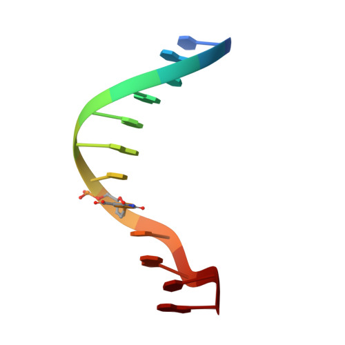
    To investigate the role of divalent cations in crystal packing, four different crystals of a Dickerson-type dodecamer with the sequence d(CGCGAATXCGCG), containing 2'-deoxy-5-formyluridine at X, were obtained under several conditions with and without divalent cations. The crystal structures are all isomorphous. The octahedrally hydrated magnesium cations found in the major groove cement the two neighbouring duplexes along the b axis. In the Mg(2+)-free crystals, a five-membered ring of water molecules occupies the same position as the magnesium site and connects the two duplexes similarly to the hydrated Mg(2+) ion. It has been concluded that water molecules can take the place of the hydrated magnesium cation in crystallization, but the magnesium cation is more effective and gives X-ray diffraction at slightly higher resolution. In all four crystals, the 5-formyluracil residues form the canonical Watson-Crick pair with adenine residues.


  • Organizational Affiliation
    • Department of Life Science, Graduate School of Bioscience and Biotechnology, Tokyo Institute of Technology, Midori-ku, Yokohama 226-8501, Japan.

Macromolecule Content 

  • Total Structure Weight: 7.38 kDa 
  • Atom Count: 675 
  • Modeled Residue Count: 24 
  • Deposited Residue Count: 24 
  • Unique nucleic acid chains: 1

Macromolecules

Find similar nucleic acids by:  Sequence
Entity ID: 1
MoleculeChains LengthOrganismImage
5'-D(*CP*GP*CP*GP*AP*AP*TP*(UFR)P*CP*GP*CP*G)-3'
A, B
12N/A
Sequence Annotations
Expand
Reference Sequence

Small Molecules

Ligands 1 Unique
IDChains Name / Formula / InChI Key2D Diagram3D Interactions
MG

Query on MG


Download:Ideal Coordinates CCD File
C [auth A]MAGNESIUM ION
Mg
JLVVSXFLKOJNIY-UHFFFAOYSA-N

Experimental Data & Validation

Experimental Data

  • Method: X-RAY DIFFRACTION
  • Resolution: 1.55 Å
  • R-Value Free:  0.232 (Depositor) 
  • R-Value Work:  0.207 (Depositor) 
Space Group: P 21 21 21
Unit Cell:
Length ( Å )Angle ( ˚ )
a = 26α = 90
b = 39.5β = 90
c = 65.7γ = 90
Software Package:
Software NamePurpose
AMoREphasing
CNSrefinement
DENZOdata reduction
SCALEPACKdata scaling

Structure Validation

View Full Validation Report


Entry History 

Deposition Data

Revision History  (Full details and data files)

  • Version 1.0: 2001-02-05
    Type: Initial release
  • Version 1.1: 2008-04-27
    Changes: Version format compliance
  • Version 1.2: 2011-07-13
    Changes: Version format compliance
  • Version 1.3: 2023-08-09
    Changes: Data collection, Database references, Derived calculations, Refinement description