1G43 | pdb_00001g43

CRYSTAL STRUCTURE OF A FAMILY IIIA CBD FROM CLOSTRIDIUM CELLULOLYTICUM


Experimental Data Snapshot

  • Method: X-RAY DIFFRACTION
  • Resolution: 2.20 Å
  • R-Value Free: 
    0.264 (Depositor), 0.264 (DCC) 
  • R-Value Work: 
    0.190 (Depositor), 0.194 (DCC) 
  • R-Value Observed: 
    0.190 (Depositor) 

Starting Model: experimental
View more details

wwPDB Validation 3D Report Full Report

Validation slider image for 1G43

This is version 1.4 of the entry. See complete history

Literature

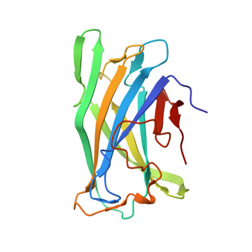
Structure of a family IIIa scaffoldin CBD from the cellulosome of Clostridium cellulolyticum at 2.2 A resolution.

Shimon, L.J.Pages, S.Belaich, A.Belaich, J.P.Bayer, E.A.Lamed, R.Shoham, Y.Frolow, F.

(2000) Acta Crystallogr D Biol Crystallogr 56: 1560-1568

  • DOI: https://doi.org/10.1107/s0907444900012889
  • Primary Citation Related Structures: 
    1G43

  • PubMed Abstract: 

    The crystal structure of the family IIIa cellulose-binding domain (CBD) from the cellulosomal scaffoldin subunit (CipC) of Clostridium cellulolyticum has been determined. The structure reveals a nine-stranded jelly-roll topology which exhibits distinctive structural elements consistent with family III CBDs that bind crystalline cellulose. These include a well conserved calcium-binding site, a putative cellulose-binding surface and a conserved shallow groove of unknown function. The CipC CBD structure is very similar to the previously elucidated family IIIa CBD from the CipA scaffoldin of C. thermocellum, with some minor differences. The CipC CBD structure was also compared with other previously described CBD structures from families IIIc and IV derived from the endoglucanases of Thermomonospora fusca and Cellulomonas fimi, respectively. The possible functional consequences of structural similarities and differences in the shallow groove and cellulose-binding faces among various CBD families and subfamilies are discussed.


  • Organizational Affiliation
    • Faculty of Chemistry, The Weizmann Institute of Science, Rehovot, Israel. linda.shimon@weizmann.ac.il

Macromolecule Content 

  • Total Structure Weight: 17.33 kDa 
  • Atom Count: 1,312 
  • Modeled Residue Count: 160 
  • Deposited Residue Count: 160 
  • Unique protein chains: 1

Macromolecules

Find similar proteins by:|  3D Structure
Entity ID: 1
MoleculeChains  Sequence LengthOrganismDetailsImage
SCAFFOLDING PROTEIN160Ruminiclostridium cellulolyticumMutation(s): 0 
UniProt
Find proteins for Q45996 (Ruminiclostridium cellulolyticum)
Explore Q45996 
Go to UniProtKB:  Q45996
Entity Groups
Sequence Clusters30% Identity50% Identity70% Identity90% Identity95% Identity100% Identity
UniProt GroupQ45996
Sequence Annotations
Expand
Reference Sequence

Experimental Data & Validation

Experimental Data

  • Method: X-RAY DIFFRACTION
  • Resolution: 2.20 Å
  • R-Value Free:  0.264 (Depositor), 0.264 (DCC) 
  • R-Value Work:  0.190 (Depositor), 0.194 (DCC) 
  • R-Value Observed: 0.190 (Depositor) 
Space Group: P 65 2 2
Unit Cell:
Length ( Å )Angle ( ˚ )
a = 46.86α = 90
b = 46.86β = 90
c = 244.121γ = 120
Software Package:
Software NamePurpose
AMoREphasing
CNSrefinement
DENZOdata reduction
SCALEPACKdata scaling

Structure Validation

View Full Validation Report


Entry History 

Deposition Data

Revision History  (Full details and data files)

  • Version 1.0: 2000-12-01
    Type: Initial release
  • Version 1.1: 2008-04-27
    Changes: Version format compliance
  • Version 1.2: 2011-07-13
    Changes: Derived calculations, Version format compliance
  • Version 1.3: 2011-11-16
    Changes: Atomic model
  • Version 1.4: 2023-08-09
    Changes: Data collection, Database references, Derived calculations, Refinement description