1G2E | pdb_00001g2e

CRYSTAL STRUCTURE OF HUD AND AU-RICH ELEMENT OF THE TUMOR NECROSIS FACTOR ALPHA RNA


Experimental Data Snapshot

  • Method: X-RAY DIFFRACTION
  • Resolution: 2.30 Å
  • R-Value Free: 
    0.238 (Depositor), 0.171 (DCC) 
  • R-Value Work: 
    0.174 (Depositor), 0.168 (DCC) 
  • R-Value Observed: 
    0.174 (Depositor) 

Starting Model: experimental
View more details

wwPDB Validation 3D Report Full Report

Validation slider image for 1G2E

This is version 1.3 of the entry. See complete history

Literature

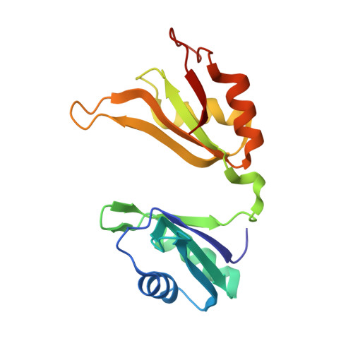
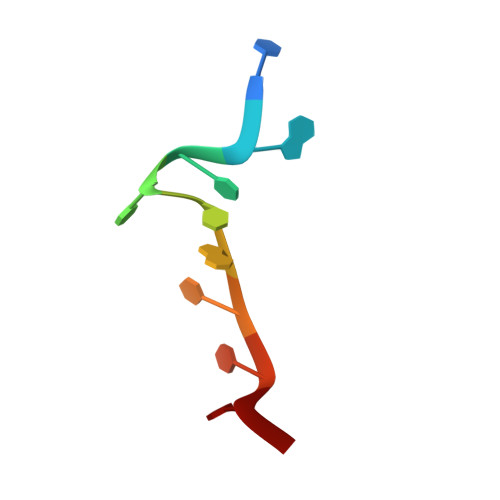
Structural basis for recognition of AU-rich element RNA by the HuD protein.

Wang, X.Tanaka Hall, T.M.

(2001) Nat Struct Biol 8: 141-145

  • DOI: https://doi.org/10.1038/84131
  • Primary Citation Related Structures: 
    1FXL, 1G2E

  • PubMed Abstract: 

    Hu proteins bind to adenosine-uridine (AU)-rich elements (AREs) in the 3' untranslated regions of many short-lived mRNAs, thereby stabilizing them. Here we report the crystal structures of the first two RNA recognition motif (RRM) domains of the HuD protein in complex with an 11-nucleotide fragment of a class I ARE (the c-fos ARE; to 1.8 A), and with an 11-nucleotide fragment of a class II ARE (the tumor necrosis factor alpha ARE; to 2.3 A). These structures reveal a consensus RNA recognition sequence that suggests a preference for pyrimidine-rich sequences and a requirement for a central uracil residue in the clustered AUUUA repeats found in class II AREs. Comparison to structures of other RRM domain-nucleic acid complexes reveals two base recognition pockets in all the structures that interact with bases using residues in conserved ribonucleoprotein motifs and at the C-terminal ends of RRM domains. Different conformations of nucleic acid can be bound by RRM domains by using different combinations of base recognition pockets and multiple RRM domains.


  • Organizational Affiliation
    • Laboratory of Structural Biology, National Institute of Environmental Health Sciences, National Institutes of Health, Research Triangle Park, North Carolina 27709, USA.

Macromolecule Content 

  • Total Structure Weight: 21.63 kDa 
  • Atom Count: 1,655 
  • Modeled Residue Count: 177 
  • Deposited Residue Count: 177 
  • Unique protein chains: 1
  • Unique nucleic acid chains: 1

Macromolecules


Find similar proteins by:|  3D Structure
Entity ID: 2
MoleculeChains  Sequence LengthOrganismDetailsImage
PARANEOPLASTIC ENCEPHALOMYELITIS ANTIGEN HUDB [auth A]167Homo sapiensMutation(s): 0 
UniProt & NIH Common Fund Data Resources
Find proteins for P26378 (Homo sapiens)
Explore P26378 
Go to UniProtKB:  P26378
PHAROS:  P26378
GTEx:  ENSG00000162374 
Entity Groups
Sequence Clusters30% Identity50% Identity70% Identity90% Identity95% Identity100% Identity
UniProt GroupP26378
Sequence Annotations
Expand
Reference Sequence
Find similar nucleic acids by:  Sequence
Entity ID: 1
MoleculeChains LengthOrganismImage
5'-R(P*UP*AP*UP*UP*UP*AP*UP*UP*UP*A)-3'A [auth B]10N/A
Sequence Annotations
Expand
Reference Sequence

Experimental Data & Validation

Experimental Data

  • Method: X-RAY DIFFRACTION
  • Resolution: 2.30 Å
  • R-Value Free:  0.238 (Depositor), 0.171 (DCC) 
  • R-Value Work:  0.174 (Depositor), 0.168 (DCC) 
  • R-Value Observed: 0.174 (Depositor) 
Space Group: P 1 21 1
Unit Cell:
Length ( Å )Angle ( ˚ )
a = 30.37α = 90
b = 100.15β = 106.59
c = 33.27γ = 90
Software Package:
Software NamePurpose
AMoREphasing
CNSrefinement
DENZOdata reduction
SCALEPACKdata scaling

Structure Validation

View Full Validation Report


Entry History 

Deposition Data

Revision History  (Full details and data files)

  • Version 1.0: 2001-02-05
    Type: Initial release
  • Version 1.1: 2008-04-27
    Changes: Version format compliance
  • Version 1.2: 2011-07-13
    Changes: Version format compliance
  • Version 1.3: 2023-08-02
    Changes: Database references, Refinement description