1G00 | pdb_00001g00

ALTERNATION OF DNA AND SOLVENT LAYERS IN THE A FORM OF D(GGCGCC) OBTAINED BY ETHANOL CRYSTALLIZATION


Experimental Data Snapshot

  • Method: X-RAY DIFFRACTION
  • Resolution: 2.30 Å
  • R-Value Free: 
    0.280 (Depositor) 
  • R-Value Work: 
    0.188 (Depositor), 0.199 (DCC) 
  • R-Value Observed: 
    0.205 (Depositor) 

Starting Model: experimental
View more details

wwPDB Validation 3D Report Full Report

Validation slider image for 1G00

This is version 1.6 of the entry. See complete history

Literature

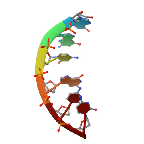
Alternation of DNA and solvent layers in the A form of d(GGCGCC) obtained by ethanol crystallization.

Urpi, L.Navaza, J.Subirana, J.A.

(2000) J Biomol Struct Dyn 18: 363-369

  • DOI: https://doi.org/10.1080/07391102.2000.10506673
  • Primary Citation Related Structures: 
    1G00

  • PubMed Abstract: 

    We have determined by X-ray crystallography the structure of the hexamer duplex d(GGCGCC)2 in the A-form using ethanol as a precipitant. The same sequence had previously been crystallized in the B-form, but with 2-methyl-2,4-pentanediol as a precipitant. It appears that ethanol precipitation is a useful method to induce the formation of A-form crystals of DNA. Packing of the molecules in the crystal has unique features: the known interaction of A-DNA duplexes between terminal base-pairs and the minor groove of neighbor molecules is combined with a superstructure consisting in an alternation of DNA layers and solvent layers (water/ions). This organization in layers has been observed before, also with hexamers in the A conformation which crystallize in the same space group (C2221). The solvent layer has a precise thickness, although very few ordered water molecules can be detected. Another feature of this crystal is its large unit cell, which gives rise to an asymmetric unit with three hexamer duplexes. One of the three duplexes is quite different from the other two in several aspects: the number of base pairs per turn, the twist pattern, the mean value of the twist angle and the fact that one terminal base-pair is not stacked as part of the duplex and appears to be disordered. So the variability in conformation of this sequence is remarkable.


  • Organizational Affiliation
    • Department d'Enginyeria Química, ETSEIB, Barcelona, Spain.

Macromolecule Content 

  • Total Structure Weight: 10.86 kDa 
  • Atom Count: 700 
  • Modeled Residue Count: 36 
  • Deposited Residue Count: 36 
  • Unique nucleic acid chains: 1

Macromolecules

Find similar nucleic acids by:  Sequence
Entity ID: 1
MoleculeChains LengthOrganismImage
5'-D(*GP*GP*CP*GP*CP*C)-3'
A, B, C, D, E
A, B, C, D, E, F
6N/A
Sequence Annotations
Expand
Reference Sequence

Experimental Data & Validation

Experimental Data

  • Method: X-RAY DIFFRACTION
  • Resolution: 2.30 Å
  • R-Value Free:  0.280 (Depositor) 
  • R-Value Work:  0.188 (Depositor), 0.199 (DCC) 
  • R-Value Observed: 0.205 (Depositor) 
Space Group: C 2 2 21
Unit Cell:
Length ( Å )Angle ( ˚ )
a = 46.149α = 90
b = 36.9β = 90
c = 110.03γ = 90
Software Package:
Software NamePurpose
AMoREphasing
SHELXL-97refinement
CAD4data reduction

Structure Validation

View Full Validation Report


Entry History 

Deposition Data

Revision History  (Full details and data files)

  • Version 1.0: 2001-02-05
    Type: Initial release
  • Version 1.1: 2008-04-27
    Changes: Version format compliance
  • Version 1.2: 2011-07-13
    Changes: Version format compliance
  • Version 1.3: 2017-10-04
    Changes: Refinement description
  • Version 1.4: 2018-04-18
    Changes: Data collection
  • Version 1.5: 2024-02-07
    Changes: Data collection, Database references
  • Version 1.6: 2024-04-03
    Changes: Refinement description