1FY2 | pdb_00001fy2

Aspartyl Dipeptidase


Experimental Data Snapshot

  • Method: X-RAY DIFFRACTION
  • Resolution: 1.20 Å
  • R-Value Free: 
    0.230 (Depositor) 
  • R-Value Work: 
    0.218 (Depositor), 0.216 (DCC) 

wwPDB Validation 3D Report Full Report

Validation slider image for 1FY2

This is version 1.4 of the entry. See complete history

Literature

The structure of aspartyl dipeptidase reveals a unique fold with a Ser-His-Glu catalytic triad.

Hakansson, K.Wang, A.H.Miller, C.G.

(2000) Proc Natl Acad Sci U S A 97: 14097-14102

  • DOI: https://doi.org/10.1073/pnas.260376797
  • Primary Citation Related Structures: 
    1FY2, 1FYE

  • PubMed Abstract: 

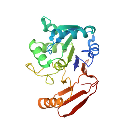
    The three-dimensional structure of Salmonella typhimurium aspartyl dipeptidase, peptidase E, was solved crystallographically and refined to 1.2-A resolution. The structure of this 25-kDa enzyme consists of two mixed beta-sheets forming a V, flanked by six alpha-helices. The active site contains a Ser-His-Glu catalytic triad and is the first example of a serine peptidase/protease with a glutamate in the catalytic triad. The active site Ser is located on a strand-helix motif reminiscent of that found in alpha/beta-hydrolases, but the polypeptide fold and the organization of the catalytic triad differ from those of the known serine proteases. This enzyme is a member of a family of serine hydrolases and appears to represent a new example of convergent evolution of peptidase activity.


  • Organizational Affiliation
    • Departments of Microbiology and Cell and Structural Biology, B103 Chemical and Life Science Laboratory, University of Illinois at Urbana-Champaign, 601 South Goodwin Avenue, Urbana, IL 61801, USA.

Macromolecule Content 

  • Total Structure Weight: 24.91 kDa 
  • Atom Count: 1,877 
  • Modeled Residue Count: 220 
  • Deposited Residue Count: 229 
  • Unique protein chains: 1

Macromolecules

Find similar proteins by:|  3D Structure
Entity ID: 1
MoleculeChains  Sequence LengthOrganismDetailsImage
ASPARTYL DIPEPTIDASE229Salmonella enterica subsp. enterica serovar TyphimuriumMutation(s): 0 
EC: 3.4 (PDB Primary Data), 3.4.13.21 (UniProt)
UniProt
Find proteins for P36936 (Salmonella typhimurium (strain LT2 / SGSC1412 / ATCC 700720))
Explore P36936 
Go to UniProtKB:  P36936
Entity Groups
Sequence Clusters30% Identity50% Identity70% Identity90% Identity95% Identity100% Identity
UniProt GroupP36936
Sequence Annotations
Expand
Reference Sequence

Small Molecules

Ligands 1 Unique
IDChains Name / Formula / InChI Key2D Diagram3D Interactions
CD

Query on CD


Download:Ideal Coordinates CCD File
B [auth A]CADMIUM ION
Cd
WLZRMCYVCSSEQC-UHFFFAOYSA-N

Experimental Data & Validation

Experimental Data

  • Method: X-RAY DIFFRACTION
  • Resolution: 1.20 Å
  • R-Value Free:  0.230 (Depositor) 
  • R-Value Work:  0.218 (Depositor), 0.216 (DCC) 
Space Group: C 1 2 1
Unit Cell:
Length ( Å )Angle ( ˚ )
a = 91.986α = 90
b = 42.69β = 106.06
c = 62.496γ = 90
Software Package:
Software NamePurpose
CNSrefinement
DENZOdata reduction
SCALEPACKdata scaling
CNSphasing

Structure Validation

View Full Validation Report


Entry History 

Deposition Data

Revision History  (Full details and data files)

  • Version 1.0: 2001-01-10
    Type: Initial release
  • Version 1.1: 2007-10-21
    Changes: Version format compliance
  • Version 1.2: 2011-07-13
    Changes: Version format compliance
  • Version 1.3: 2011-11-16
    Changes: Atomic model
  • Version 1.4: 2024-02-07
    Changes: Data collection, Database references, Derived calculations