1FWI | pdb_00001fwi

KLEBSIELLA AEROGENES UREASE, H134A VARIANT


Experimental Data Snapshot

  • Method: X-RAY DIFFRACTION
  • Resolution: 2.00 Å
  • R-Value Work: 
    0.170 (Depositor), 0.162 (DCC) 
  • R-Value Observed: 
    0.170 (Depositor) 

wwPDB Validation 3D Report Full Report

Validation slider image for 1FWI

This is version 1.3 of the entry. See complete history

Literature

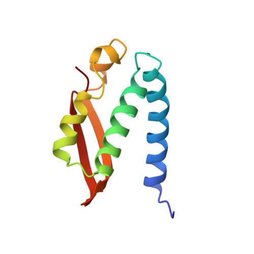
Characterization of the mononickel metallocenter in H134A mutant urease.

Park, I.S.Michel, L.O.Pearson, M.A.Jabri, E.Karplus, P.A.Wang, S.Dong, J.Scott, R.A.Koehler, B.P.Johnson, M.K.Hausinger, R.P.

(1996) J Biological Chem 271: 18632-18637

  • DOI: https://doi.org/10.1074/jbc.271.31.18632
  • Primary Citation Related Structures: 
    1FWI

  • PubMed Abstract: 

    A mutant form of Klebsiella aerogenes urease possessing Ala instead of His at position 134 (H134A) is inactive and binds approximately half the normal complement of nickel (Park, I.-S., and Hausinger, R. P.(1993) Protein Sci. 2, 1034-1041). The crystal structure of the H134A protein was obtained at 2.0-A resolution, and it confirms that only Ni-1 of the two nickel ions found in the native enzyme is present. In contrast to the pseudotetrahedral geometry observed for Ni-1 in native urease (where it is liganded by His-246, His-272, one oxygen atom of carbamylated Lys-217, and a water molecule at partial occupancy), the mononickel metallocenter in the H134A protein was found to possess octahedral geometry and was coordinated by the above protein ligands plus three water molecules. The nickel site of H134A urease was probed by UV-visible, variable temperature magnetic circular dichroism, and x-ray absorption spectroscopies. The spectroscopic data are consistent with the presence of Ni(II) in octahedral geometry coordinated by two histidylimidazoles and additional oxygen and/or nitrogen donors. These data underscore the requirement of Ni-2 for formation of active urease and demonstrate the important role of Ni-2 in establishing the proper Ni-1 coordination geometry.


  • Organizational Affiliation
    • Department of Microbiology, Michigan State University, East Lansing, Michigan 48824-1101, USA.

Macromolecule Content 

  • Total Structure Weight: 83.21 kDa 
  • Atom Count: 5,979 
  • Modeled Residue Count: 752 
  • Deposited Residue Count: 773 
  • Unique protein chains: 3

Macromolecules

Find similar proteins by:|  3D Structure
Entity ID: 1
MoleculeChains  Sequence LengthOrganismDetailsImage
UREASE100Klebsiella aerogenesMutation(s): 0 
EC: 3.5.1.5
UniProt
Find proteins for P18316 (Klebsiella aerogenes)
Explore P18316 
Go to UniProtKB:  P18316
Entity Groups
Sequence Clusters30% Identity50% Identity70% Identity90% Identity95% Identity100% Identity
UniProt GroupP18316
Sequence Annotations
Expand
Reference Sequence
Find similar proteins by:|  3D Structure
Entity ID: 2
MoleculeChains  Sequence LengthOrganismDetailsImage
UREASE106Klebsiella aerogenesMutation(s): 0 
EC: 3.5.1.5
UniProt
Find proteins for P18315 (Klebsiella aerogenes)
Explore P18315 
Go to UniProtKB:  P18315
Entity Groups
Sequence Clusters30% Identity50% Identity70% Identity90% Identity95% Identity100% Identity
UniProt GroupP18315
Sequence Annotations
Expand
Reference Sequence
Find similar proteins by:|  3D Structure
Entity ID: 3
MoleculeChains  Sequence LengthOrganismDetailsImage
UREASE567Klebsiella aerogenesMutation(s): 2 
EC: 3.5.1.5
UniProt
Find proteins for P18314 (Klebsiella aerogenes)
Explore P18314 
Go to UniProtKB:  P18314
Entity Groups
Sequence Clusters30% Identity50% Identity70% Identity90% Identity95% Identity100% Identity
UniProt GroupP18314
Sequence Annotations
Expand
Reference Sequence

Small Molecules

Ligands 1 Unique
IDChains Name / Formula / InChI Key2D Diagram3D Interactions
NI

Query on NI


Download:Ideal Coordinates CCD File
D [auth C]NICKEL (II) ION
Ni
VEQPNABPJHWNSG-UHFFFAOYSA-N
Modified Residues  1 Unique
IDChains TypeFormula2D DiagramParent
KCX
Query on KCX
C
L-PEPTIDE LINKINGC7 H14 N2 O4LYS

Experimental Data & Validation

Experimental Data

  • Method: X-RAY DIFFRACTION
  • Resolution: 2.00 Å
  • R-Value Work:  0.170 (Depositor), 0.162 (DCC) 
  • R-Value Observed: 0.170 (Depositor) 
Space Group: I 21 3
Unit Cell:
Length ( Å )Angle ( ˚ )
a = 170.8α = 90
b = 170.8β = 90
c = 170.8γ = 90
Software Package:
Software NamePurpose
X-PLORmodel building
X-PLORrefinement
DENZOdata reduction
SCALEPACKdata scaling
X-PLORphasing

Structure Validation

View Full Validation Report


Entry History 

Deposition Data

Revision History  (Full details and data files)

  • Version 1.0: 1997-10-15
    Type: Initial release
  • Version 1.1: 2008-03-24
    Changes: Version format compliance
  • Version 1.2: 2011-07-13
    Changes: Derived calculations, Version format compliance
  • Version 1.3: 2021-11-03
    Changes: Database references, Derived calculations, Other