1FP4 | pdb_00001fp4

CRYSTAL STRUCTURE OF THE ALPHA-H195Q MUTANT OF NITROGENASE


Experimental Data Snapshot

  • Method: X-RAY DIFFRACTION
  • Resolution: 2.50 Å
  • R-Value Free: 
    0.240 (Depositor) 
  • R-Value Work: 
    0.184 (Depositor) 

wwPDB Validation 3D Report Full Report

Validation slider image for 1FP4

This is version 1.5 of the entry. See complete history

Literature

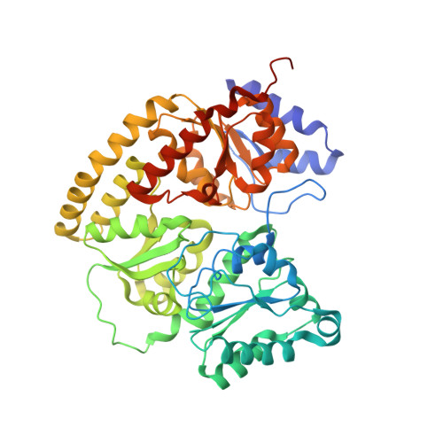
Mechanistic features and structure of the nitrogenase alpha-Gln195 MoFe protein

Sorlie, M.Christiansen, J.Lemon, B.J.Peters, J.W.Dean, D.R.Hales, B.J.

(2001) Biochemistry 40: 1540-1549

  • DOI: https://doi.org/10.1021/bi0013997
  • Primary Citation Related Structures: 
    1FP4

  • PubMed Abstract: 

    EPR signals observed under CO and C(2)H(2) during nitrogenase turnover were investigated for the alpha-Gln(195) MoFe protein, an altered form for which the alpha-His(195) residue has been substituted by glutamine. Under CO, samples show S = 1/2 hi- and lo-CO EPR signals identical to those recognized for the wild-type protein, whereas the S = 3/2 signals generated under high CO/high flux conditions differ. Previous work has revealed that the EPR spectrum generated under C(2)H(2) exhibits a signal (S(EPR1)) originating from the FeMo-cofactor having two or more bound C(2)H(2) adducts and a second signal (S(EPR2)) arising from a radical species [Sørlie, M., Christiansen, J., Dean, D. R., and Hales, B. J. (1999) J. Am. Chem. Soc. 121, 9457-9458]. Pressure-dependent studies show that the intensity of these signals has a sigmoidal dependency at low pressures and maximized at 0.1 atm C(2)H(2) with a subsequent decrease in steady-state intensity at higher pressures. Analogous signals are not recognized for the wild-type MoFe protein. Analysis of the principal g-factors of S(EPR2) suggests that it either represents an unusual metal cluster or is a carboxylate centered radical possibly originating from homocitrate. Both S(EPR1) and S(EPR2) exhibit similar relaxation properties that are atypical for S = 1/2 signals originating from Fe-S clusters or radicals and indicate a coupled relaxation pathway. The alpha-Gln(195) MoFe protein also exhibits these signals when incubated under turnover conditions in the presence of C(2)H(4). Under these conditions, additional inflections in the g 4-6 region assigned to ground-state transitions of an S = 3/2 spin system are also recognized and assigned to turnover states of the MoFe protein without C(2)H(4) bound. The structure of alpha-Gln(195) was crystallographically determined and found to be virtually identical to that of the wild-type MoFe protein except for replacement of an NuH-S hydrogen bond interaction between FeMo-cofactor and the imidazole side chain of alpha-His(195) by an analogous interaction involving Gln.


  • Organizational Affiliation
    • Department of Chemistry, Louisiana State University, Baton Rouge, LA 70803-1804, USA.

Macromolecule Content 

  • Total Structure Weight: 233.23 kDa 
  • Atom Count: 16,295 
  • Modeled Residue Count: 1,979 
  • Deposited Residue Count: 2,030 
  • Unique protein chains: 2

Macromolecules

Find similar proteins by:|  3D Structure
Entity ID: 1
MoleculeChains  Sequence LengthOrganismDetailsImage
NITROGENASE MOLYBDENUM-IRON PROTEIN ALPHA CHAIN
A, C
492Azotobacter vinelandiiMutation(s): 1 
EC: 1.18.6.1
UniProt
Find proteins for P07328 (Azotobacter vinelandii)
Explore P07328 
Go to UniProtKB:  P07328
Entity Groups
Sequence Clusters30% Identity50% Identity70% Identity90% Identity95% Identity100% Identity
UniProt GroupP07328
Sequence Annotations
Expand
Reference Sequence
Find similar proteins by:|  3D Structure
Entity ID: 2
MoleculeChains  Sequence LengthOrganismDetailsImage
NITROGENASE MOLYBDENUM-IRON PROTEIN BETA CHAIN
B, D
523Azotobacter vinelandiiMutation(s): 0 
EC: 1.18.6.1
UniProt
Find proteins for P07329 (Azotobacter vinelandii)
Explore P07329 
Go to UniProtKB:  P07329
Entity Groups
Sequence Clusters30% Identity50% Identity70% Identity90% Identity95% Identity100% Identity
UniProt GroupP07329
Sequence Annotations
Expand
Reference Sequence

Small Molecules

Ligands 4 Unique
IDChains Name / Formula / InChI Key2D Diagram3D Interactions
CFM

Query on CFM


Download:Ideal Coordinates CCD File
F [auth A],
J [auth C]
FE-MO-S CLUSTER
Fe7 Mo S9
UZRXIPMKRKMLQF-UHFFFAOYSA-N
CLP

Query on CLP


Download:Ideal Coordinates CCD File
G [auth A],
K [auth C]
FE-S CLUSTER
Fe8 S8
MJXAKWFPOPRKKV-UHFFFAOYSA-N
HCA

Query on HCA


Download:Ideal Coordinates CCD File
E [auth A],
I [auth C]
3-HYDROXY-3-CARBOXY-ADIPIC ACID
C7 H10 O7
XKJVEVRQMLKSMO-SSDOTTSWSA-N
CA

Query on CA


Download:Ideal Coordinates CCD File
H [auth B],
L [auth D]
CALCIUM ION
Ca
BHPQYMZQTOCNFJ-UHFFFAOYSA-N

Experimental Data & Validation

Experimental Data

  • Method: X-RAY DIFFRACTION
  • Resolution: 2.50 Å
  • R-Value Free:  0.240 (Depositor) 
  • R-Value Work:  0.184 (Depositor) 
Space Group: P 1 21 1
Unit Cell:
Length ( Å )Angle ( ˚ )
a = 107.2α = 90
b = 130.2β = 111.2
c = 80.4γ = 90
Software Package:
Software NamePurpose
SCALEPACKdata scaling
AMoREphasing
X-PLORrefinement

Structure Validation

View Full Validation Report


Entry History 

Deposition Data

Revision History  (Full details and data files)

  • Version 1.0: 2002-11-20
    Type: Initial release
  • Version 1.1: 2008-04-27
    Changes: Version format compliance
  • Version 1.2: 2011-07-13
    Changes: Version format compliance
  • Version 1.3: 2017-10-04
    Changes: Refinement description
  • Version 1.4: 2021-11-10
    Changes: Database references, Derived calculations
  • Version 1.5: 2024-05-29
    Changes: Data collection