1FNO | pdb_00001fno

PEPTIDASE T (TRIPEPTIDASE)


Experimental Data Snapshot

  • Method: X-RAY DIFFRACTION
  • Resolution: 2.40 Å
  • R-Value Free: 
    0.261 (Depositor) 
  • R-Value Work: 
    0.222 (Depositor), 0.220 (DCC) 

wwPDB Validation 3D Report Full Report

Validation slider image for 1FNO

This is version 1.3 of the entry. See complete history

Literature

Structure of Peptidase T from Salmonella typhimurium

Hakansson, K.Miller, C.G.

(2002) Eur J Biochem 269: 443-450

  • DOI: https://doi.org/10.1046/j.0014-2956.2001.02665.x
  • Primary Citation Related Structures: 
    1FNO

  • PubMed Abstract: 

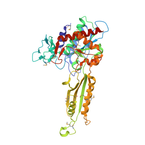
    The structure of peptidase T, or tripeptidase, was determined by multiple wavelength anomalous dispersion (MAD) methodology and refined to 2.4 A resolution. Peptidase T comprises two domains; a catalytic domain with an active site containing two metal ions, and a smaller domain formed through a long insertion into the catalytic domain. The two metal ions, presumably zinc, are separated by 3.3 A, and are coordinated by five carboxylate and histidine ligands. The molecular surface of the active site is negatively charged. Peptidase T has the same basic fold as carboxypeptidase G2. When the structures of the two enzymes are superimposed, a number of homologous residues, not evident from the sequence alone, could be identified. Comparison of the active sites of peptidase T, carboxypeptidase G2, Aeromonas proteolytica aminopeptidase, carboxypeptidase A and leucine aminopeptidase reveals a common structural framework with interesting similarities and differences in the active sites and in the zinc coordination. A putative binding site for the C-terminal end of the tripeptide substrate was found at a peptidase T specific fingerprint sequence motif.


  • Organizational Affiliation
    • Department of Microbiology, University of Illinois at Urbana-Champaign, Urbana, IL 61801, USA.

Macromolecule Content 

  • Total Structure Weight: 47 kDa 
  • Atom Count: 3,260 
  • Modeled Residue Count: 408 
  • Deposited Residue Count: 417 
  • Unique protein chains: 1

Macromolecules

Find similar proteins by:|  3D Structure
Entity ID: 1
MoleculeChains  Sequence LengthOrganismDetailsImage
PEPTIDASE T417Salmonella enterica subsp. enterica serovar TyphimuriumMutation(s): 15 
EC: 3.4.11 (PDB Primary Data), 3.4.11.4 (UniProt)
UniProt
Find proteins for P26311 (Salmonella typhimurium (strain LT2 / SGSC1412 / ATCC 700720))
Explore P26311 
Go to UniProtKB:  P26311
Entity Groups
Sequence Clusters30% Identity50% Identity70% Identity90% Identity95% Identity100% Identity
UniProt GroupP26311
Sequence Annotations
Expand
Reference Sequence

Experimental Data & Validation

Experimental Data

  • Method: X-RAY DIFFRACTION
  • Resolution: 2.40 Å
  • R-Value Free:  0.261 (Depositor) 
  • R-Value Work:  0.222 (Depositor), 0.220 (DCC) 
Space Group: C 1 2 1
Unit Cell:
Length ( Å )Angle ( ˚ )
a = 132.374α = 90
b = 46.036β = 116.11
c = 96.591γ = 90
Software Package:
Software NamePurpose
CNSrefinement
DENZOdata reduction
SCALEPACKdata scaling
CNSphasing

Structure Validation

View Full Validation Report


Entry History 

Deposition Data

Revision History  (Full details and data files)

  • Version 1.0: 2002-03-13
    Type: Initial release
  • Version 1.1: 2008-04-27
    Changes: Version format compliance
  • Version 1.2: 2011-07-13
    Changes: Derived calculations, Version format compliance
  • Version 1.3: 2024-11-13
    Changes: Data collection, Database references, Derived calculations, Structure summary