1FNE | pdb_00001fne

HISTOCOMPATIBILITY ANTIGEN


Experimental Data Snapshot

  • Method: X-RAY DIFFRACTION
  • Resolution: 1.90 Å
  • R-Value Free: 
    0.270 (Depositor) 
  • R-Value Work: 
    0.231 (Depositor) 
  • R-Value Observed: 
    0.231 (Depositor) 

Starting Model: experimental
View more details

wwPDB Validation 3D Report Full Report

Validation slider image for 1FNE

This is version 1.7 of the entry. See complete history

Literature

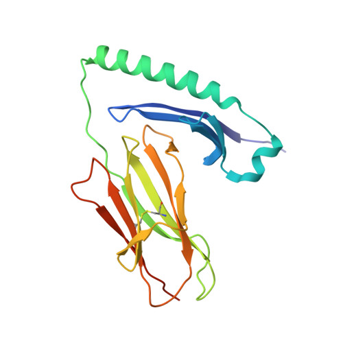
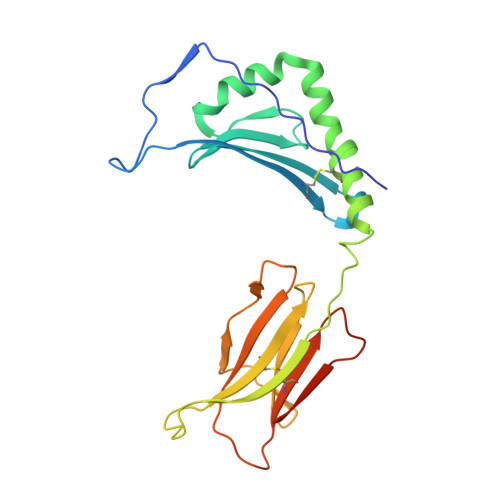
Structural and functional consequences of altering a peptide MHC anchor residue.

Kersh, G.J.Miley, M.J.Nelson, C.A.Grakoui, A.Horvath, S.Donermeyer, D.L.Kappler, J.Allen, P.M.Fremont, D.H.

(2001) J Immunol 166: 3345-3354

  • DOI: https://doi.org/10.4049/jimmunol.166.5.3345
  • Primary Citation Related Structures: 
    1FNE, 1FNG

  • PubMed Abstract: 

    To better understand TCR discrimination of multiple ligands, we have analyzed the crystal structures of two Hb peptide/I-E(k) complexes that differ by only a single amino acid substitution at the P6 anchor position within the peptide (E73D). Detailed comparison of multiple independently determined structures at 1.9 A resolution reveals that removal of a single buried methylene group can alter a critical portion of the TCR recognition surface. Significant variance was observed in the peptide P5-P8 main chain as well as a rotamer difference at LeuP8, approximately 10 A distal from the substitution. No significant variations were observed in the conformation of the two MHC class II molecules. The ligand alteration results in two peptide/MHC complexes that generate bulk T cell responses that are distinct and essentially nonoverlapping. For the Hb-specific T cell 3.L2, substitution reduces the potency of the ligand 1000-fold. Soluble 3.L2 TCR binds the two peptide/MHC complexes with similar affinity, although with faster kinetics. These results highlight the role of subtle variations in MHC Ag presentation on T cell activation and signaling.


  • Organizational Affiliation
    • Department of Pathology and Center for Immunology, Washington University School of Medicine, St. Louis, MO 63110, USA.

Macromolecule Content 

  • Total Structure Weight: 96.77 kDa 
  • Atom Count: 7,011 
  • Modeled Residue Count: 790 
  • Deposited Residue Count: 832 
  • Unique protein chains: 2

Macromolecules

Find similar proteins by:|  3D Structure
Entity ID: 1
MoleculeChains  Sequence LengthOrganismDetailsImage
PROTEIN (MHC CLASS II I-EK, ALPHA CHAIN)
A, C
192Mus musculusMutation(s): 0 
UniProt
Find proteins for P04224 (Mus musculus)
Explore P04224 
Go to UniProtKB:  P04224
Entity Groups
Sequence Clusters30% Identity50% Identity70% Identity90% Identity95% Identity100% Identity
UniProt GroupP04224
Sequence Annotations
Expand
Reference Sequence
Find similar proteins by:|  3D Structure
Entity ID: 2
MoleculeChains  Sequence LengthOrganismDetailsImage
PROTEIN (MHC CLASS II I-EK, BETA CHAIN)
B, D
224Mus musculusMutation(s): 1 
UniProt
Find proteins for P02089 (Mus musculus)
Explore P02089 
Go to UniProtKB:  P02089
Entity Groups
Sequence Clusters30% Identity50% Identity70% Identity90% Identity95% Identity100% Identity
UniProt GroupP02089
Sequence Annotations
Expand
Reference Sequence

Experimental Data & Validation

Experimental Data

  • Method: X-RAY DIFFRACTION
  • Resolution: 1.90 Å
  • R-Value Free:  0.270 (Depositor) 
  • R-Value Work:  0.231 (Depositor) 
  • R-Value Observed: 0.231 (Depositor) 
Space Group: C 1 2 1
Unit Cell:
Length ( Å )Angle ( ˚ )
a = 146.9α = 90
b = 56.853β = 91.76
c = 116.726γ = 90
Software Package:
Software NamePurpose
DENZOdata reduction
SCALEPACKdata scaling
AMoREphasing
CNSrefinement

Structure Validation

View Full Validation Report


Entry History 

Deposition Data

Revision History  (Full details and data files)

  • Version 1.0: 2001-03-07
    Type: Initial release
  • Version 1.1: 2007-10-16
    Changes: Version format compliance
  • Version 1.2: 2011-07-13
    Changes: Derived calculations, Version format compliance
  • Version 1.3: 2017-08-23
    Changes: Advisory, Refinement description, Source and taxonomy
  • Version 1.4: 2018-03-14
    Changes: Database references
  • Version 1.5: 2020-07-29
    Type: Remediation
    Reason: Carbohydrate remediation
    Changes: Data collection, Database references, Derived calculations, Structure summary
  • Version 1.6: 2023-08-09
    Changes: Advisory, Data collection, Database references, Refinement description, Structure summary
  • Version 1.7: 2024-11-20
    Changes: Structure summary