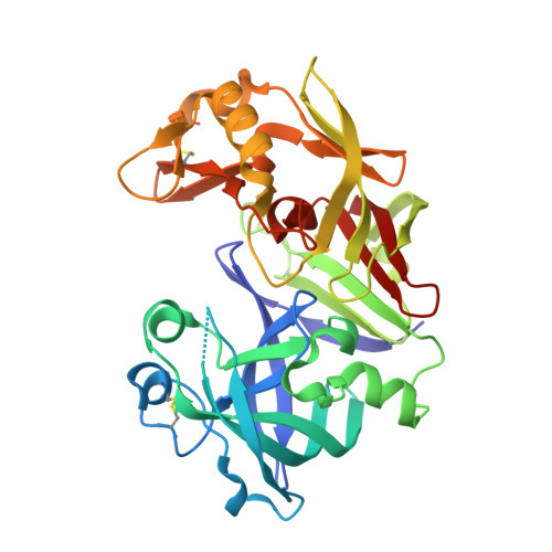
1FMX

STRUCTURE OF NATIVE PROTEINASE A IN THE SPACE GROUP P21


Experimental Data Snapshot

  • Method: X-RAY DIFFRACTION
  • Resolution: 2.61 Å
  • R-Value Free: 0.279 
  • R-Value Work: 0.205 
  • R-Value Observed: 0.205 

wwPDB Validation   3D Report Full Report


Ligand Structure Quality Assessment 


This is version 2.1 of the entry. See complete history


Literature

An unusual orientation for Tyr75 in the active site of the aspartic proteinase from Saccharomyces cerevisiae.

Gustchina, A.Li, M.Phylip, L.H.Lees, W.E.Kay, J.Wlodawer, A.

(2002) Biochem Biophys Res Commun 295: 1020-1026

  • DOI: https://doi.org/10.1016/s0006-291x(02)00742-8
  • Primary Citation of Related Structures:  
    1FMU, 1FMX

  • PubMed Abstract: 

    The structures of the native Saccharomyces cerevisiae proteinase A have been solved by molecular replacement in the monoclinic and trigonal crystal forms and refined at 2.6-2.7A resolution. These structures agree overall with those of other uninhibited aspartic proteinases. However, an unusual orientation for the side chain of Tyr75, a conserved residue on the flexible "flap" that covers the active site and is important for the activity of these enzymes, was found in the trigonal crystals. A similar conformation of Tyr75 occupying the S1 substrate-binding pocket was previously reported only for chymosin (where it was interpreted as representing a "self-inhibited" state of the enzyme), but for no other aspartic proteinases. Since this orientation of Tyr75 has now been seen in the structures of two members of the family of aspartic proteinases, it might indicate that the placement of that residue in the S1 substrate-binding pocket might have some functional significance, analogous to what was seen for self-inhibited structures of serine proteinases.


  • Organizational Affiliation

    Protein Structure Section, Macromolecular Crystallography Laboratory, National Cancer Institute at Frederick, Frederick, MD 21702-1201, USA.


Macromolecules
Oligosaccharides

Help

Entity ID: 2
MoleculeChains Length2D Diagram Glycosylation3D Interactions
beta-D-mannopyranose-(1-4)-2-acetamido-2-deoxy-beta-D-glucopyranose-(1-4)-2-acetamido-2-deoxy-beta-D-glucopyranose
C
3N-Glycosylation
Glycosylation Resources
GlyTouCan:  G15407YE
GlyCosmos:  G15407YE
GlyGen:  G15407YE
Entity ID: 3
MoleculeChains Length2D Diagram Glycosylation3D Interactions
2-acetamido-2-deoxy-beta-D-glucopyranose-(1-4)-2-acetamido-2-deoxy-beta-D-glucopyranose
D
2N-Glycosylation
Glycosylation Resources
GlyTouCan:  G42666HT
GlyCosmos:  G42666HT
GlyGen:  G42666HT
Small Molecules
Ligands 1 Unique
IDChains Name / Formula / InChI Key2D Diagram3D Interactions
NAG
Query on NAG

Download Ideal Coordinates CCD File 
E [auth A],
F [auth B]
2-acetamido-2-deoxy-beta-D-glucopyranose
C8 H15 N O6
OVRNDRQMDRJTHS-FMDGEEDCSA-N
Experimental Data & Validation

Experimental Data

  • Method: X-RAY DIFFRACTION
  • Resolution: 2.61 Å
  • R-Value Free: 0.279 
  • R-Value Work: 0.205 
  • R-Value Observed: 0.205 
  • Space Group: P 1 21 1
Unit Cell:
Length ( Å )Angle ( ˚ )
a = 82.964α = 90
b = 49.074β = 96.5
c = 94.685γ = 90
Software Package:
Software NamePurpose
SCALEPACKdata scaling
AMoREphasing
CNSrefinement

Structure Validation

View Full Validation Report


Ligand Structure Quality Assessment 


Entry History 

Deposition Data

Revision History  (Full details and data files)

  • Version 1.0: 2002-07-31
    Type: Initial release
  • Version 1.1: 2008-04-27
    Changes: Version format compliance
  • Version 1.2: 2011-07-13
    Changes: Non-polymer description, Version format compliance
  • Version 1.3: 2017-10-04
    Changes: Advisory, Refinement description
  • Version 2.0: 2020-07-29
    Type: Remediation
    Reason: Carbohydrate remediation
    Changes: Atomic model, Data collection, Derived calculations, Structure summary
  • Version 2.1: 2024-10-30
    Changes: Advisory, Data collection, Database references, Structure summary