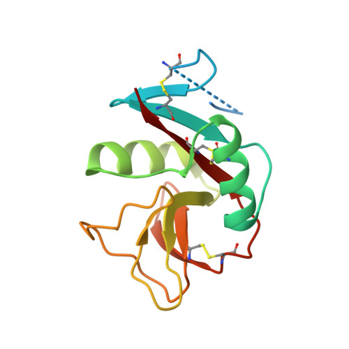
Crystal structure of human CD69: a C-type lectin-like activation marker of hematopoietic cells.
Natarajan, K., Sawicki, M.W., Margulies, D.H., Mariuzza, R.A.(2000) Biochemistry 39: 14779-14786
- PubMed: 11101293
- DOI: https://doi.org/10.1021/bi0018180
- Primary Citation of Related Structures:
1FM5 - PubMed Abstract:
CD69 is a widely expressed type II transmembrane glycoprotein related to the C-type animal lectins that exhibits regulated expression on a variety of cells of the hematopoietic lineage, including neutrophils, monocytes, T cells, B cells, natural killer (NK) cells, and platelets. Activation of T lymphocytes results in the induced expression of CD69 at the cell surface. In addition, cross-linking of CD69 by specific antibodies leads to the activation of cells bearing this receptor and to the induction of effector functions. However, the physiological ligand of CD69 is unknown. We report here the X-ray crystal structure of the extracellular C-type lectin-like domain (CTLD) of human CD69 at 2.27 A resolution. Recombinant CD69 was expressed in bacterial inclusion bodies and folded in vitro. The protein, which exists as a disulfide-linked homodimer on the cell surface, crystallizes as a symmetrical dimer, similar to those formed by the related NK cell receptors Ly49A and CD94. The structure reveals conservation of the C-type lectin-like fold, including preservation of the two alpha-helical regions found in Ly49A and mannose-binding protein (MBP). However, only one of the nine residues coordinated to Ca(2+) in MBP is conserved in CD69 and no bound Ca(2+) is evident in the crystal structure. Surprisingly, electron density suggestive of a puckered six-membered ring was discovered at a site structurally analogous to the ligand-binding sites of MBP and Ly49A. This sugar-like density may represent, or mimic, part of the natural ligand recognized by CD69.
- Molecular Biology Section, Laboratory of Immunology, National Institute of Allergy and Infectious Diseases, National Institutes of Health, Bethesda, Maryland 20892, USA.
Organizational Affiliation:


