1FL3 | pdb_00001fl3

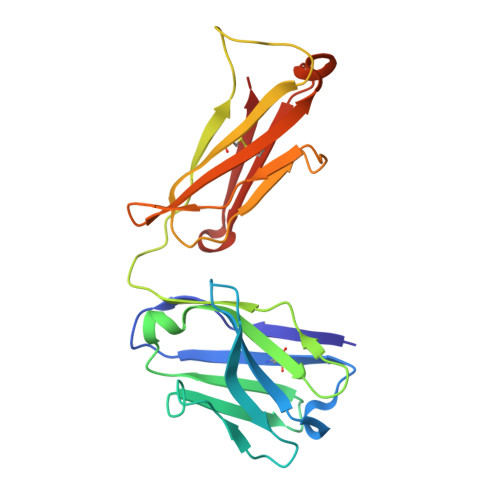
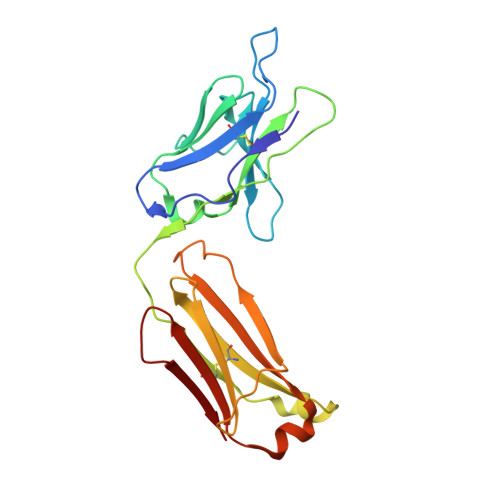
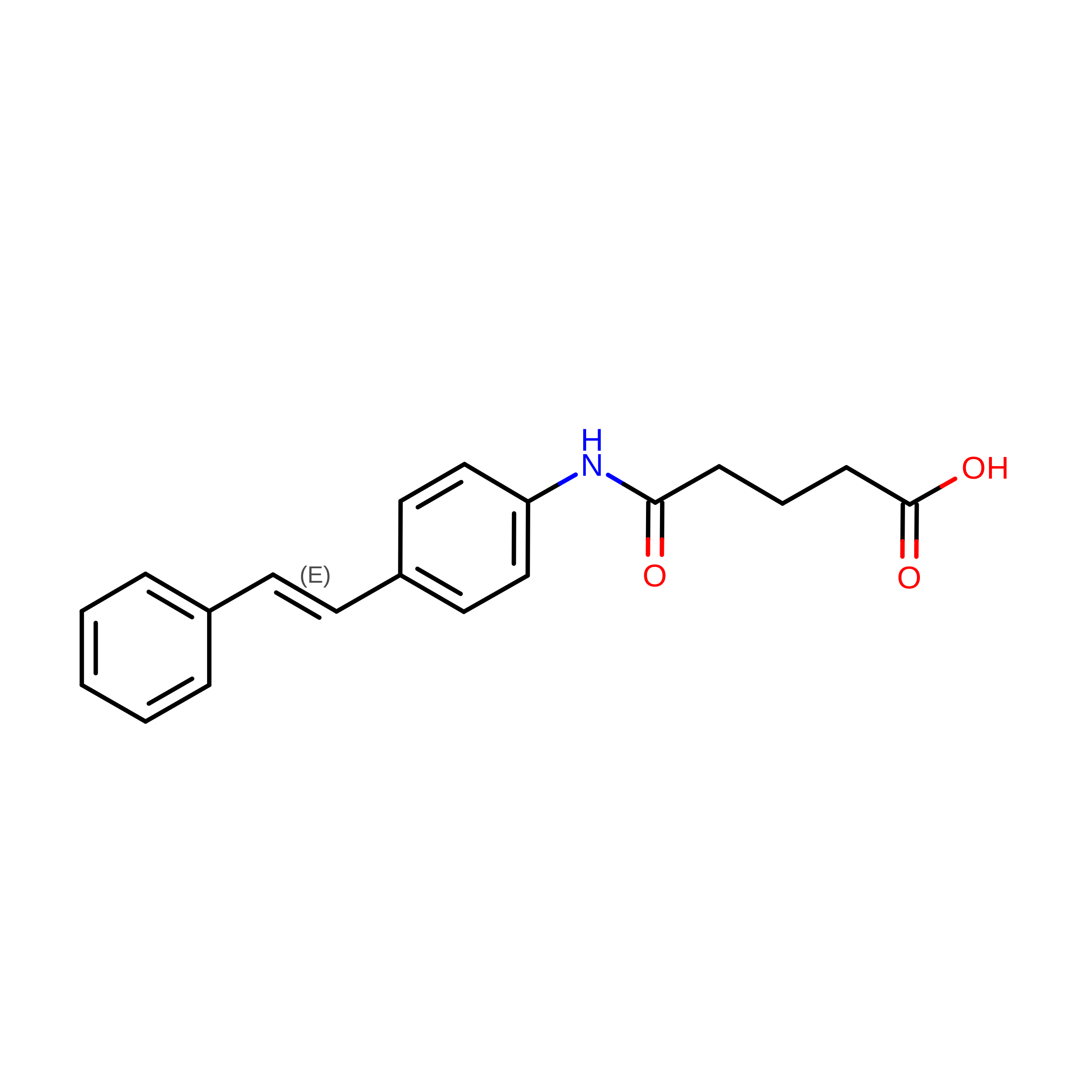
CRYSTAL STRUCTURE OF THE BLUE FLUORESCENT ANTIBODY (19G2) IN COMPLEX WITH STILBENE HAPTEN AT 277K


Experimental Data Snapshot

  • Method: X-RAY DIFFRACTION
  • Resolution: 2.45 Å
  • R-Value Free: 
    0.262 (Depositor) 
  • R-Value Work: 
    0.231 (Depositor), 0.240 (DCC) 
  • R-Value Observed: 
    0.100 (Depositor) 

wwPDB Validation 3D Report Full Report

Validation slider image for 1FL3

Ligand Structure Quality Assessment 


This is version 1.5 of the entry. See complete history. 

Literature

Blue-fluorescent antibodies.

Simeonov, A., Matsushita, M., Juban, E.A., Thompson, E.H., Hoffman, T.Z., Beuscher IV, A.E., Taylor, M.J., Wirsching, P., Rettig, W., McCusker, J.K., Stevens, R.C., Millar, D.P., Schultz, P.G., Lerner, R.A., Janda, K.D.

(2000) Science 290: 307-313

  • DOI: https://doi.org/10.1126/science.290.5490.307
  • Primary Citation Related Structures: 
    1FL3

  • PubMed Abstract: 

    The forte of catalytic antibodies has resided in the control of the ground-state reaction coordinate. A principle and method are now described in which antibodies can direct the outcome of photophysical and photochemical events that take place on excited-state potential energy surfaces. The key component is a chemically reactive optical sensor that provides a direct report of the dynamic interplay between protein and ligand at the active site. To illustrate the concept, we used a trans-stilbene hapten to elicit a panel of monoclonal antibodies that displayed a range of fluorescent spectral behavior when bound to a trans-stilbene substrate. Several antibodies yielded a blue fluorescence indicative of an excited-state complex or "exciplex" between trans-stilbene and the antibody. The antibodies controlled the isomerization coordinate of trans-stilbene and dynamically coupled this manifold with an active-site residue. A step was taken toward the use of antibody-based photochemical sensors for diagnostic and clinical applications.


  • Organizational Affiliation: 
    • Department of Chemistry, The Scripps Research Institute and the Skaggs Institute for Chemical Biology, 10550 North Torrey Pines Road, La Jolla, CA 92037, USA.

Macromolecule Content 

  • Total Structure Weight: 91.18 kDa 
  • Atom Count: 6,633 
  • Modeled Residue Count: 844 
  • Deposited Residue Count: 844 
  • Unique protein chains: 2

Macromolecules

Find similar proteins by:|  3D Structure
Entity ID: 1
MoleculeChains  Sequence LengthOrganismDetailsImage
BLUE FLUORESCENT ANTIBODY (19G2)-HEAVY CHAINA [auth H],
C [auth A]
208Mus musculusMutation(s): 0 
UniProt
Find proteins for P01867 (Mus musculus)
Explore P01867 
Go to UniProtKB:  P01867
Entity Groups
Sequence Clusters30% Identity50% Identity70% Identity90% Identity95% Identity100% Identity
UniProt GroupP01867
Sequence Annotations
Expand
Reference Sequence
Find similar proteins by:|  3D Structure
Entity ID: 2
MoleculeChains  Sequence LengthOrganismDetailsImage
BLUE FLUORESCENT ANTIBODY (19G2)-LIGHT CHAINB [auth L],
D [auth B]
214Mus musculusMutation(s): 0 
UniProt
Find proteins for P01837 (Mus musculus)
Explore P01837 
Go to UniProtKB:  P01837
Entity Groups
Sequence Clusters30% Identity50% Identity70% Identity90% Identity95% Identity100% Identity
UniProt GroupP01837
Sequence Annotations
Expand
Reference Sequence

Experimental Data & Validation

Experimental Data

  • Method: X-RAY DIFFRACTION
  • Resolution: 2.45 Å
  • R-Value Free:  0.262 (Depositor) 
  • R-Value Work:  0.231 (Depositor), 0.240 (DCC) 
  • R-Value Observed: 0.100 (Depositor) 
Space Group: C 1 2 1
Unit Cell:
Length ( Å )Angle ( ˚ )
a = 196.93α = 90
b = 62.069β = 117.54
c = 93.539γ = 90
Software Package:
Software NamePurpose
DENZOdata reduction
SCALEPACKdata scaling
MOLREPphasing
CNSrefinement

Structure Validation

View Full Validation Report


Ligand Structure Quality Assessment 


Entry History 

Deposition Data

Revision History  (Full details and data files)

  • Version 1.0: 2000-11-01
    Type: Initial release
  • Version 1.1: 2008-04-27
    Changes: Version format compliance
  • Version 1.2: 2011-07-13
    Changes: Version format compliance
  • Version 1.3: 2017-10-11
    Changes: Data collection
  • Version 1.4: 2018-01-31
    Changes: Database references
  • Version 1.5: 2024-10-30
    Changes: Data collection, Database references, Derived calculations, Structure summary