1FAO | pdb_00001fao

STRUCTURE OF THE PLECKSTRIN HOMOLOGY DOMAIN FROM DAPP1/PHISH IN COMPLEX WITH INOSITOL 1,3,4,5-TETRAKISPHOSPHATE


Experimental Data Snapshot

  • Method: X-RAY DIFFRACTION
  • Resolution: 1.80 Å
  • R-Value Free: 
    0.232 (Depositor) 
  • R-Value Work: 
    0.219 (Depositor) 

Starting Model: experimental
View more details

wwPDB Validation 3D Report Full Report

Validation slider image for 1FAO

This is version 1.4 of the entry. See complete history

Literature

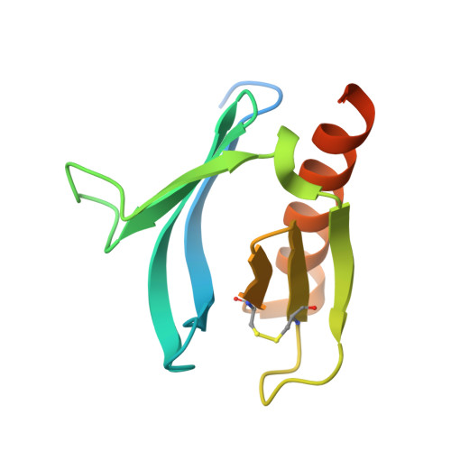
Structural basis for discrimination of 3-phosphoinositides by pleckstrin homology domains.

Ferguson, K.M.Kavran, J.M.Sankaran, V.G.Fournier, E.Isakoff, S.J.Skolnik, E.Y.Lemmon, M.A.

(2000) Mol Cell 6: 373-384

  • DOI: https://doi.org/10.1016/s1097-2765(00)00037-x
  • Primary Citation Related Structures: 
    1FAO, 1FB8, 1FHW, 1FHX

  • PubMed Abstract: 

    Pleckstrin homology (PH) domains are protein modules of around 120 amino acids found in many proteins involved in cellular signaling. Certain PH domains drive signal-dependent membrane recruitment of their host proteins by binding strongly and specifically to lipid second messengers produced by agonist-stimulated phosphoinositide 3-kinases (PI 3-Ks). We describe X-ray crystal structures of two different PH domains bound to Ins(1,3,4,5)P4, the head group of the major PI 3-K product PtdIns(3,4,5)P3. One of these PH domains (from Grp1) is PtdIns(3,4,5)P3 specific, while the other (from DAPP1/PHISH) binds strongly to both PtdIns(3,4,5)P3 and its 5'-dephosphorylation product, PtdIns(3,4)P2. Comparison of the two structures provides an explanation for the distinct phosphoinositide specificities of the two PH domains and allows us to predict the 3-phosphoinositide selectivity of uncharacterized PH domains.


  • Organizational Affiliation
    • Department of Biochemistry and Biophysics and The Johnson Foundation, University of Pennsylvania School of Medicine, Philadelphia 19104, USA.

Macromolecule Content 

  • Total Structure Weight: 15.31 kDa 
  • Atom Count: 971 
  • Modeled Residue Count: 100 
  • Deposited Residue Count: 126 
  • Unique protein chains: 1

Macromolecules

Find similar proteins by:|  3D Structure
Entity ID: 1
MoleculeChains  Sequence LengthOrganismDetailsImage
DUAL ADAPTOR OF PHOSPHOTYROSINE AND 3-PHOSPHOINOSITIDES126Homo sapiensMutation(s): 0 
Membrane Entity: Yes 
UniProt & NIH Common Fund Data Resources
Find proteins for Q9UN19 (Homo sapiens)
Explore Q9UN19 
Go to UniProtKB:  Q9UN19
PHAROS:  Q9UN19
GTEx:  ENSG00000070190 
Entity Groups
Sequence Clusters30% Identity50% Identity70% Identity90% Identity95% Identity100% Identity
UniProt GroupQ9UN19
Sequence Annotations
Expand
Reference Sequence

Small Molecules

Ligands 1 Unique
IDChains Name / Formula / InChI Key2D Diagram3D Interactions
4IP

Query on 4IP


Download:Ideal Coordinates CCD File
B [auth A]INOSITOL-(1,3,4,5)-TETRAKISPHOSPHATE
C6 H16 O18 P4
CIPFCGZLFXVXBG-CNWJWELYSA-N

Experimental Data & Validation

Experimental Data

  • Method: X-RAY DIFFRACTION
  • Resolution: 1.80 Å
  • R-Value Free:  0.232 (Depositor) 
  • R-Value Work:  0.219 (Depositor) 
Space Group: P 21 21 21
Unit Cell:
Length ( Å )Angle ( ˚ )
a = 40.674α = 90
b = 46.273β = 90
c = 64.743γ = 90
Software Package:
Software NamePurpose
DENZOdata reduction
SCALEPACKdata scaling
AMoREphasing
CNSrefinement

Structure Validation

View Full Validation Report


Entry History 

Deposition Data

Revision History  (Full details and data files)

  • Version 1.0: 2000-07-20
    Type: Initial release
  • Version 1.1: 2008-04-27
    Changes: Version format compliance
  • Version 1.2: 2011-07-13
    Changes: Version format compliance
  • Version 1.3: 2023-08-09
    Changes: Data collection, Database references, Derived calculations, Refinement description
  • Version 1.4: 2024-10-30
    Changes: Structure summary